15 Endocrine System
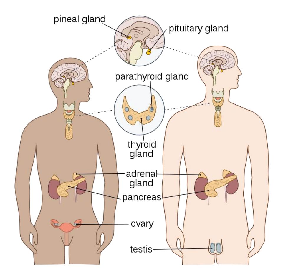
Figure 15.1 Human Endocrine System with Labels.
15.0.1 Endocrine System
15.0.2 Terms
- Pituitary
- Anterior pituitary gland
- Posterior pituitary gland
- Hypothalamus
- Pineal body/gland
- Thyroid gland
- Parathyroid glands
15.1 Pancreas
- Pancreatic Islets (Islets of Langerhans)
- Alpha cells (α cells)
- Beta cells (β-cells)
- Adrenal gland
- Adrenal cortex
- Adrenal medulla
- Testis
- Ovary
15.2 Prelab Activities
15.3 Prelab Activity 15.1
15.4 Definitions
- Endocrine System __________________________________________________
- Anterior pituitary gland ___________________________________________
- Posterior pituitary gland __________________________________________
- Hypothalamus _________________________________________________
- Pineal body/gland ______________________________________________
- Thyroid gland __________________________________________________
- Parathyroid glands ______________________________________________
- Pancreas _____________________________________________________
- Pancreatic Islets (Islets of Langerhans) ______________________________________________________________
- Alpha cells (α cells) _____________________________________________
- Beta cells ( β-cells) _____________________________________________
- Adrenal gland _________________________________________________
- Adrenal cortex _________________________________________________
- Adrenal medulla _______________________________________________
- Testis ________________________________________________________
- Ovary ________________________________________________________
15.5 Prelab Activity 15.2
15.6 Identifications
Label the following components of the endocrine system: 15.2 – 15.4
Label the components A, B and C.
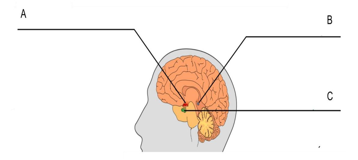
Figure 15.2 Midsagittal Section of the Human Brain.
Identify the structure of the endocrine organ listed below.
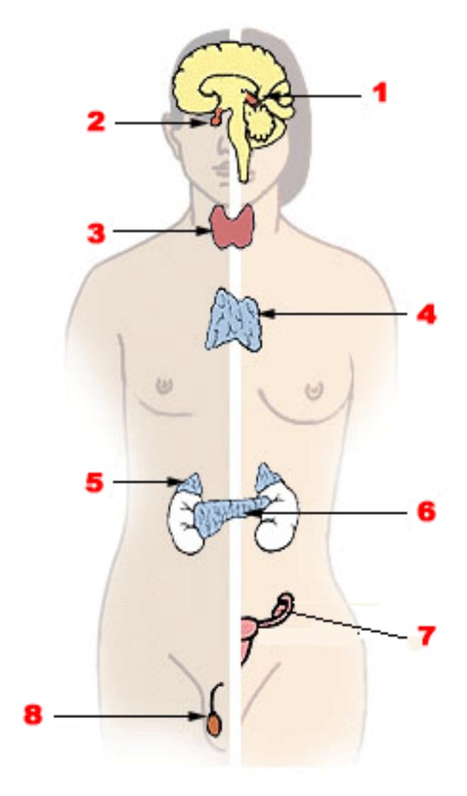
Figure 15.3 Endocrine System Illustration.
| 1. | 5. |
| 2. | 6. |
| 3. | 7. |
| 4. | 8. |
Label structures and cells designated by 1, 2, and the arrow.
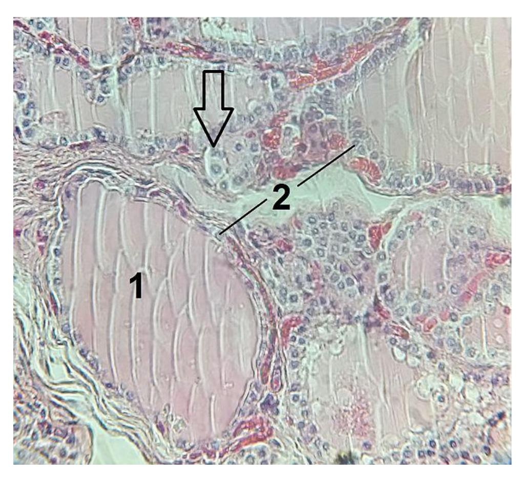
Figure 15.4 Histology of the Thyroid Gland.
- #1 structure: ___________________________________________
- #2 (cell): ______________________________________________
- Arrow➡️: (cell) ___________________________________________
15.7 Lab Activities
15.8 Endocrine System
15.8.1 Terms
Pituitary
- Anterior pituitary gland
- Posterior pituitary gland
Hypothalamus
Pineal body/gland
Thyroid gland
Parathyroid glands
Pancreas
Adrenal gland
- Adrenal cortex
- Adrenal medulla
Reproductive glands
- Testis
- Ovary
15.9 Lab Activity 15.1:
From the models’ storage area obtain a plastic model of the human brain and an endocrine system HubbardTM Scientific model.
- Identify and label the structures and regions listed above.
- Feel the structures you are identifying. Provide a list of the hormones released by the endocrine organs and include their physiological function.
- Then take a picture for your later reference.
- Have another group, or your instructor review your demonstrations.
- If time permits, create a five-minute review training video of the structures noted above.
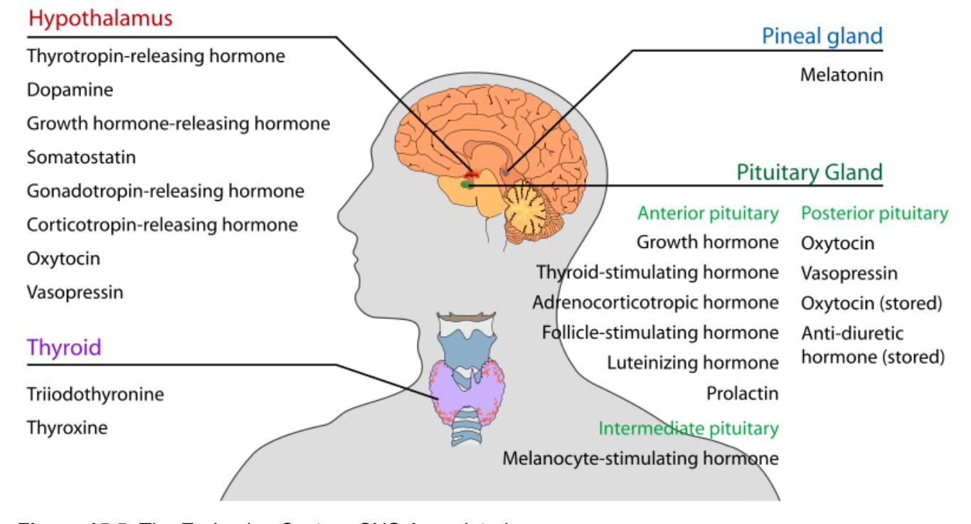
Figure 15.5 The Endocrine System-CNS Associated.
15.9.1 The Hypothalamus- Pituitary complex
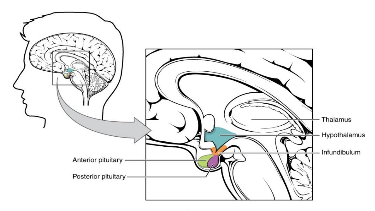
Figure 15.6 The Hypothalamus-Pituitary Complex.
15.9.2 The Pituitary Regions
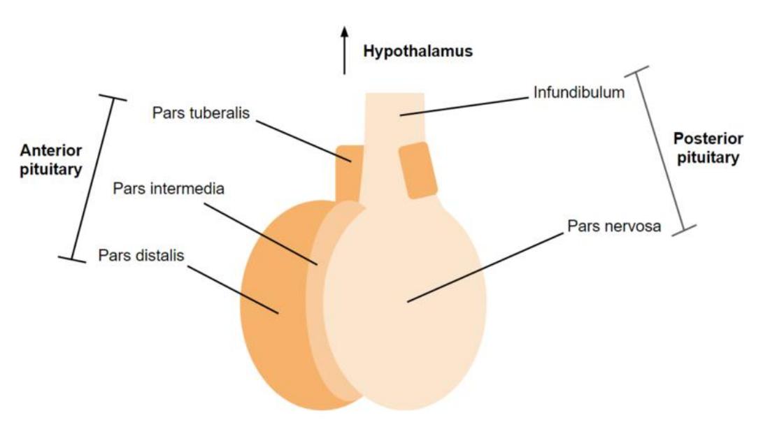
Figure 15.7 Pituitary Gland Diagram.
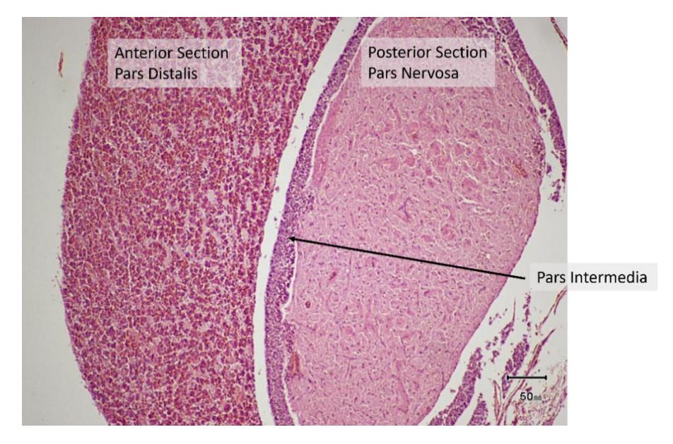
Figure 15.8 Pituitary Photomicrograph Showing the Anterior and Posterior Sections.
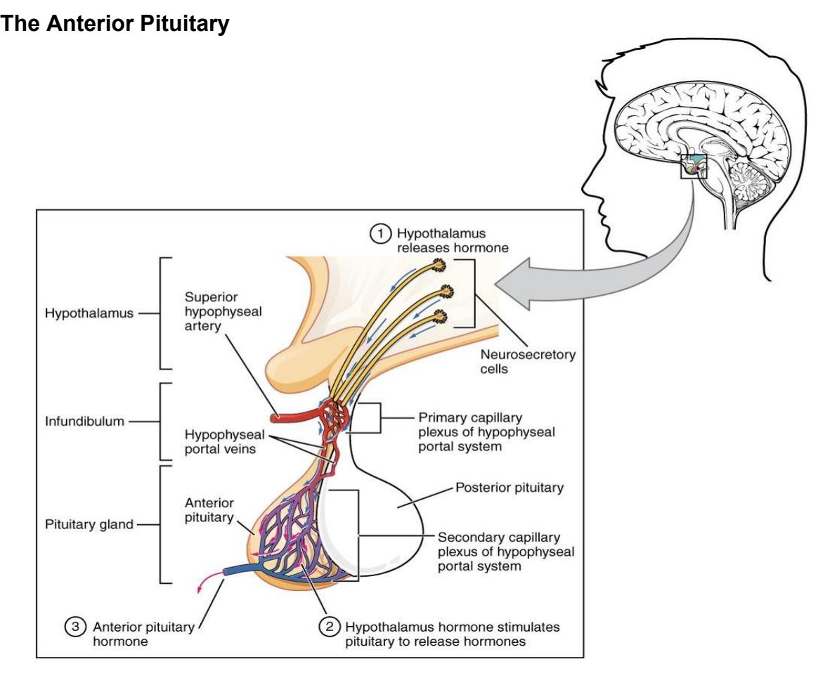
Figure 15.9 The Anterior Pituitary Complex.
15.10 Anterior Pituitary Hormones
Figure 15.10 Hormones of the Pituitary Complex, Anterior Section.
15.11 The Posterior Pituitary
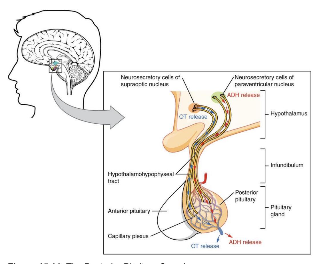
Figure 15.11 The Posterior Pituitary Complex.
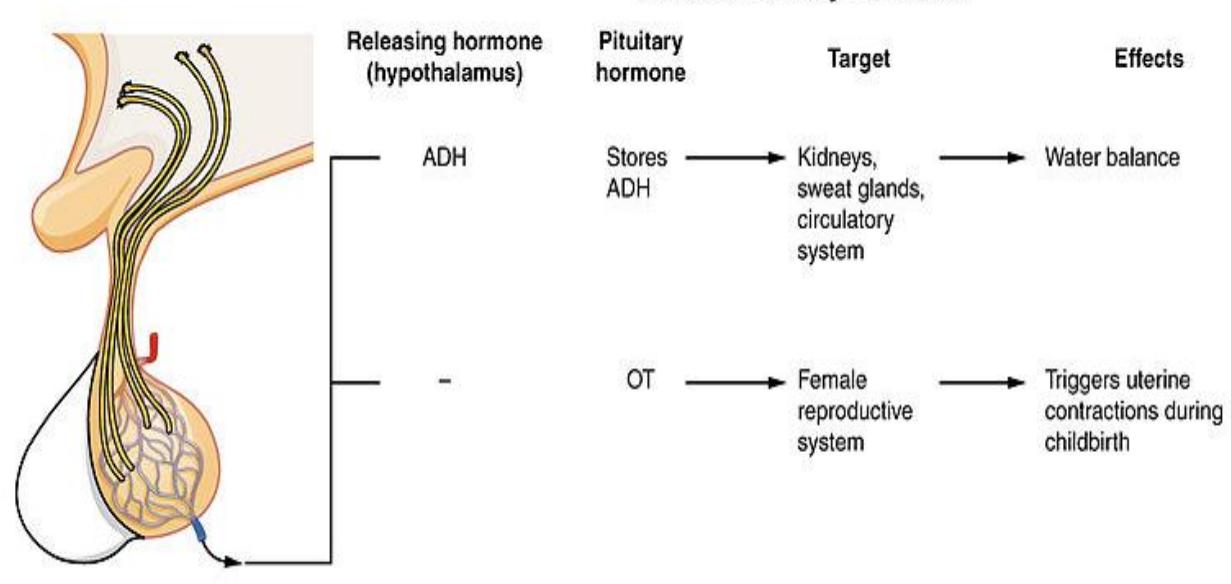
Figure 15.12 Hormones of the Pituitary Complex, Posterior Section.
15.12 Pineal gland
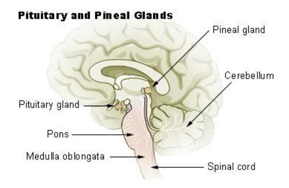
Figure 15.13 Mid-Sagittal Section of the Human Brain; Pituitary and Pineal Glands.
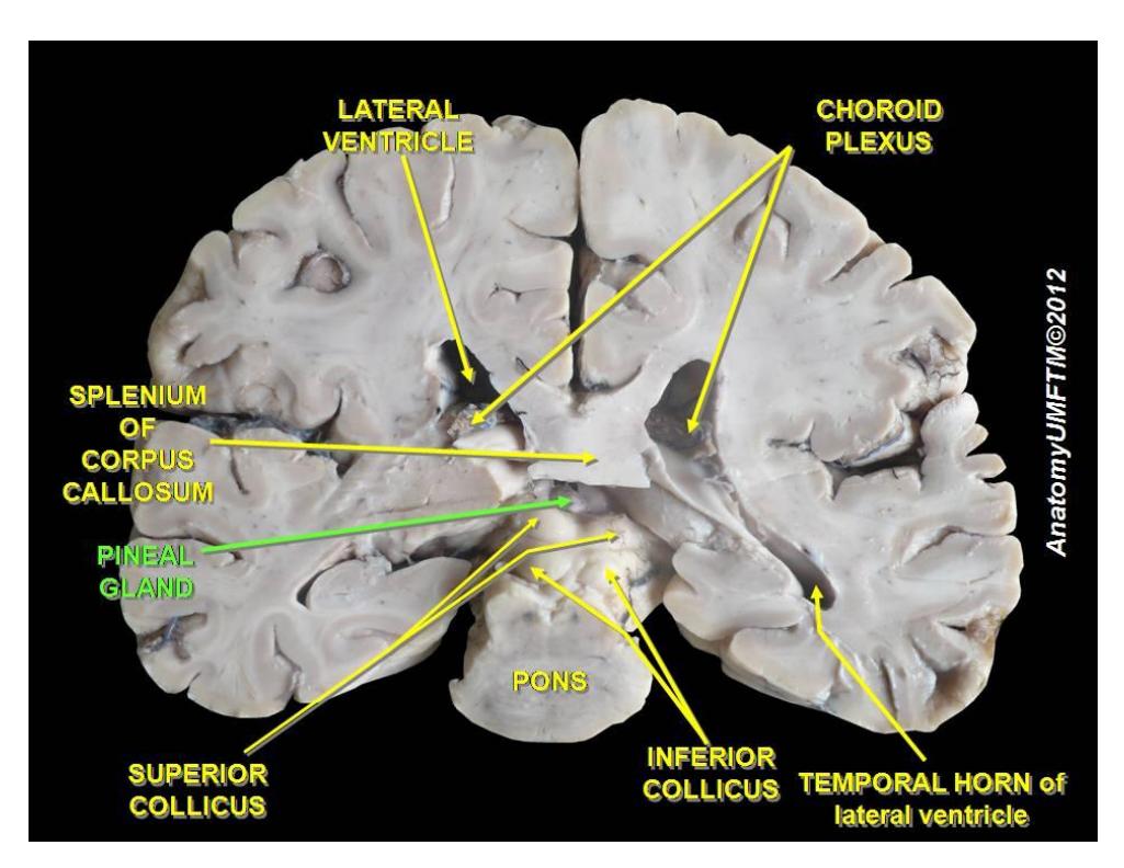
Figure 15.14 Coronal Dissection of the Human Brain Highlighting the Pineal Gland.
15.13 Thyroid and Parathyroid Glands
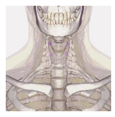
Figure 15.15 Parathyroid and Thyroid Glands in Color
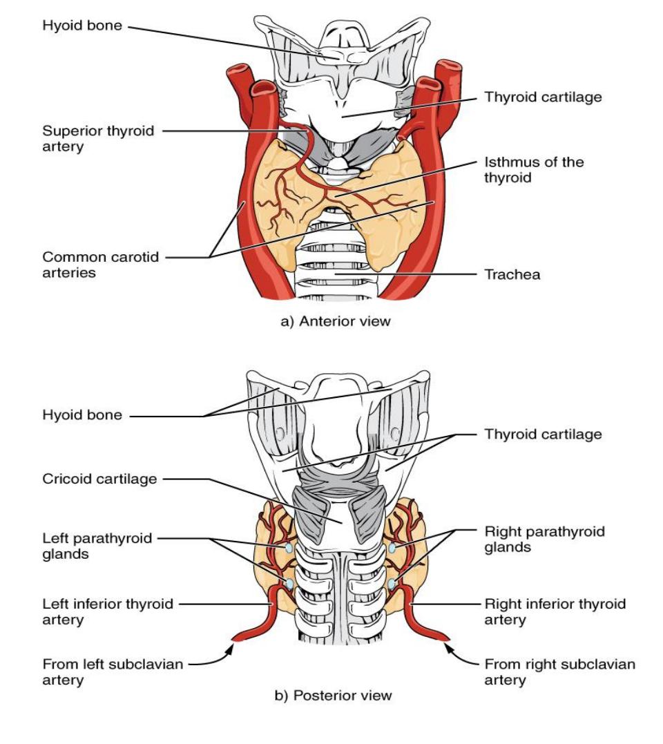
Figure 15.16 Thyroid and Parathyroid Glands.
15.14 Parathyroid Gland
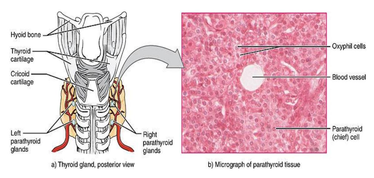
Figure 15.17 Parathyroid Glands.
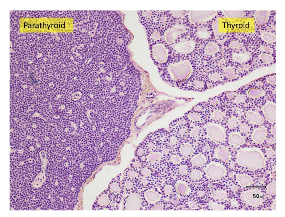
Figure 15.18 Photomicrograph of the Thyroid and Parathyroid Glands.
15.15 Pancreas
15.15.1 Terms
- Endocrine cells; Pancreatic Islets (Islets of Langerhans)
- Alpha cells (α-cells)
- Beta cells (β-cells)
- Exocrine cells
- Acinar cells
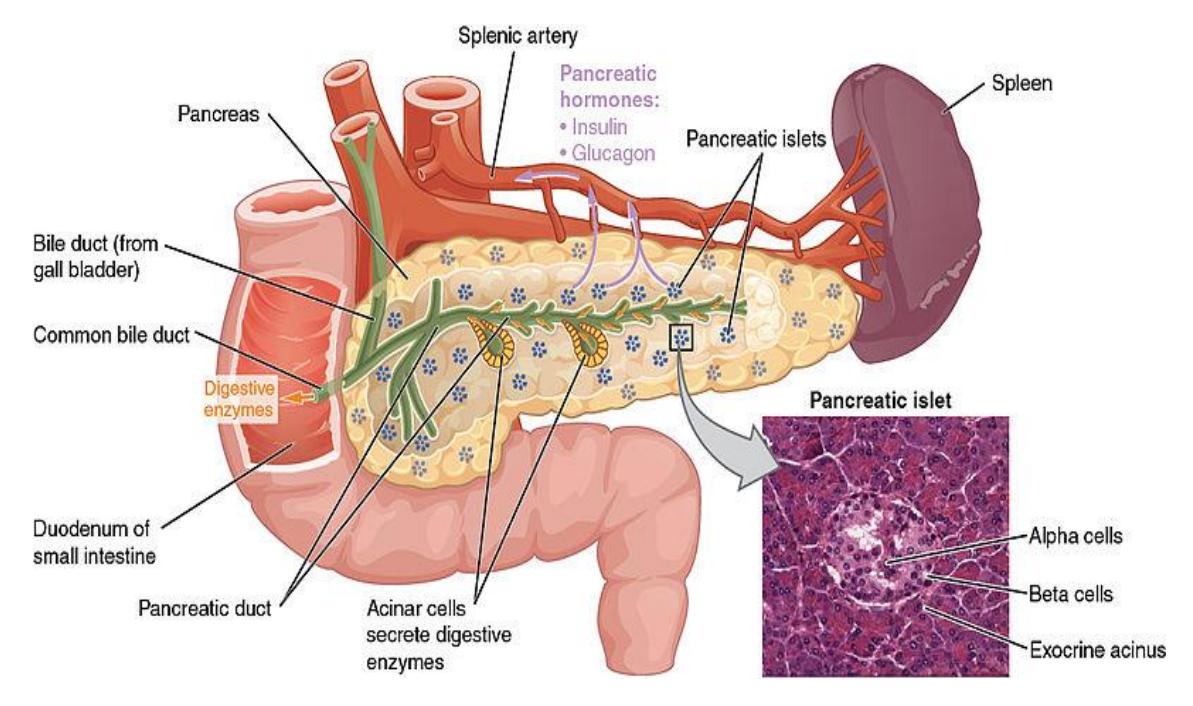
Figure 15.19. The pancreas.
15.15.2 Adrenal glands - (on the superior margin of each kidney)
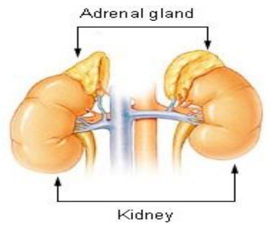
Figure 15.20 Adrenal Glands.
15.15.3 Adrenal gland
- Adrenal cortex
- Adrenal medulla
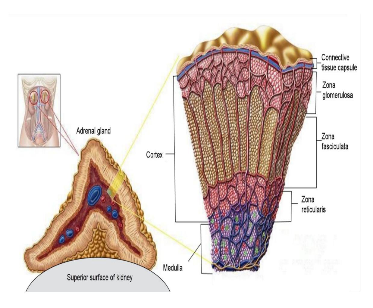
Figure 15.21 Depiction of the Adrenal Gland.
15.16 Cross section of an adrenal gland
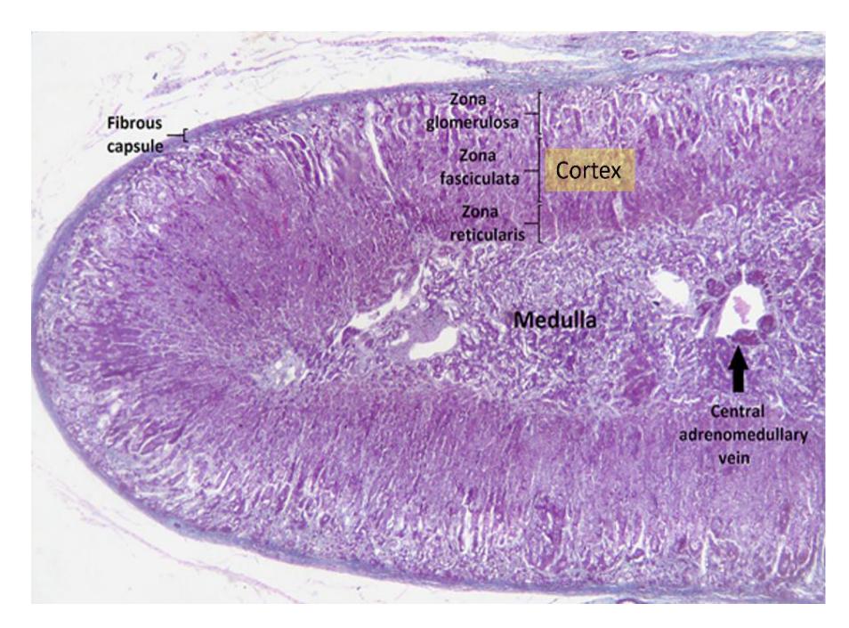
Figure 15.22 Photomicrograph of a Human Adrenal Gland.
15.16.1 Hormones of the Adrenal Glands
- Adrenal cortex (releases corticosteroid hormones)
- Zona glomerulosa: aldosterone (mineralocorticoid) retention of sodium and excretion of potassium (sodium retention increases water and blood volume and blood pressure)
- Zona fasciculata: cortisol (glucocorticoid) (hydrocortisone) increases blood glucose, also depresses inflammatory and allergic responses.
- Zona reticularis: androgens supplement male hormones
- adrenal medulla 80% epinephrine and 20% norepinephrine (stimulated by sympathetic fibers) - brings about sympathetic effects.
15.16.2 Lab Activity 15.2: (optional upon the discretion of the Instructor)
From the microscope storage area obtain a microscope and a slide of the adrenal gland. In a group of two, focus on the adrenal tissue at 400x.
- Identify and label the structures and regions listed above.
- Provide a list of the hormones released by the cortex and medulla and include their physiological function.
- Then take a picture for your later reference.
- Have another group, or your instructor review your demonstrations.
- If time permits, create a five-minute review training video of the structures noted above.
15.17 Reproductive Organs
15.18 Testes and Ovaries
15.18.1 Terms
- Testicle
- Ovary
These organs may be covered in detail in a later section by your Instructor.
15.18.2 Testes
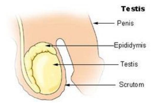
Figure 15.23 Testis.
15.18.3 Ovaries
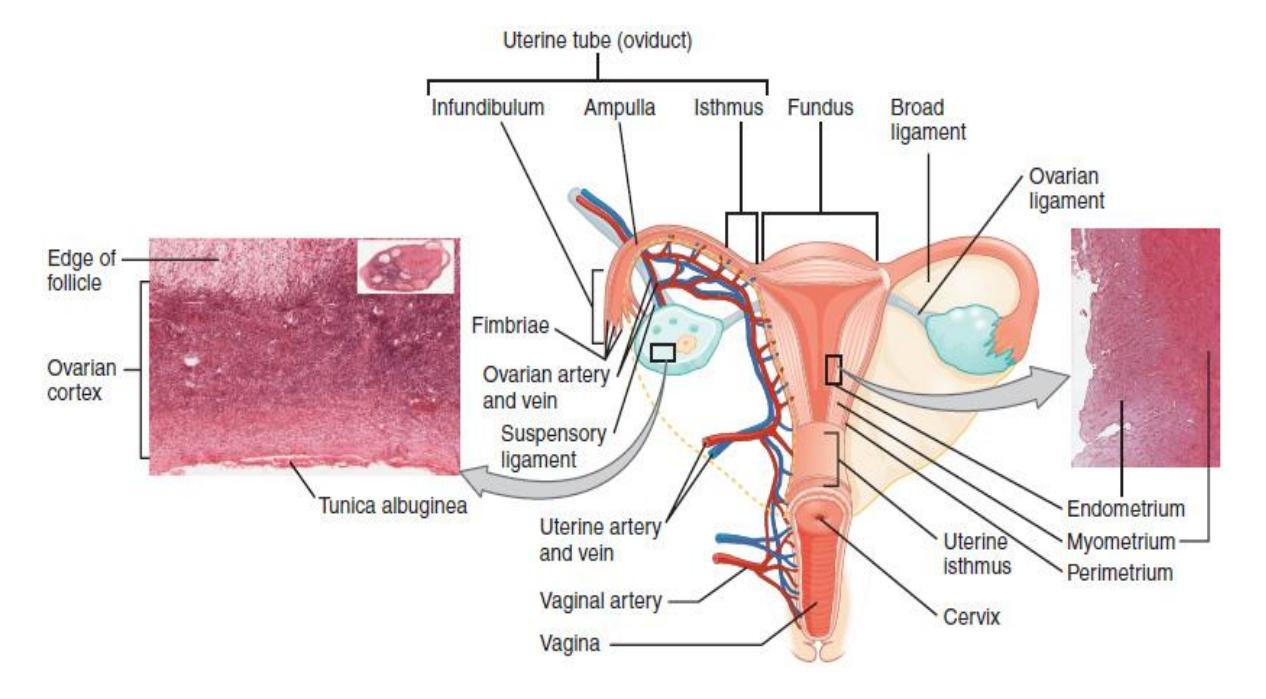
Figure 15.24 The Reproductive System, Female.
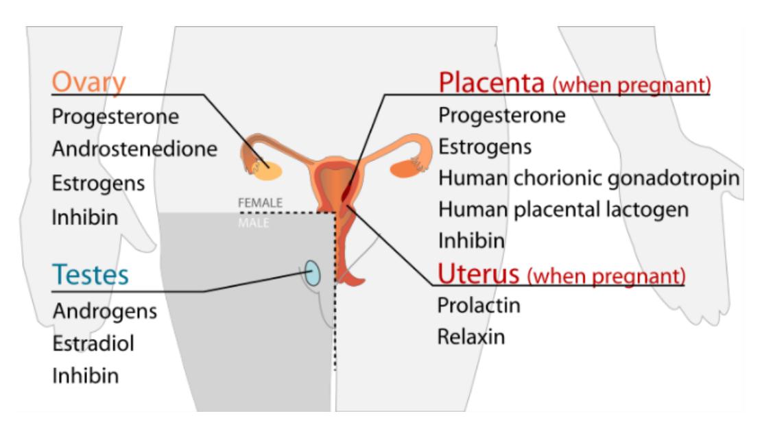
Figure 15.25 List of the Hormones Found in the Endocrine Glands of the Reproductive System.
15.19 Lab Activity 15.3: (45 minutes)
From the microscope and slide storage area(s) obtain a microscope and a set of slides of each of the following endocrine organs:
- Pituitary gland
- Anterior pituitary gland
- Posterior pituitary gland
- Thyroid gland
- Parathyroid glands
- Pancreas
- Focus on a magnification of 400X on each of the tissues.
- Identify the tissues and regions listed above as instructed by your instructor.
- Take a picture of the tissues with your cell phone. And download them onto your computer. Label the tissues and provide a list of the hormones released by the endocrine organs and include their physiological function.
- Have your instructor review your identification.
- After the class, create a review training video of the tissues and structures noted above.
15.19.1 Examples of the Endocrine Tissues
15.20 Pituitary Tissues: Anterior and Posterior Aspects
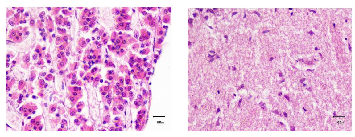
Figure 15.26 The Pituitary: a) Anterior Pituitary on the Left and b) the Posterior Pituitary on the Right.
15.20.1 Thyroid and Parathyroid Glands
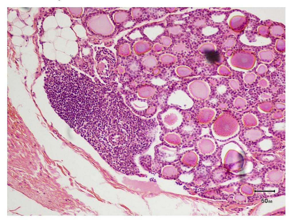
Figure 15.27 Micrograph of the Thyroid and Parathyroid Glands.
15.21 Pancreas tissue
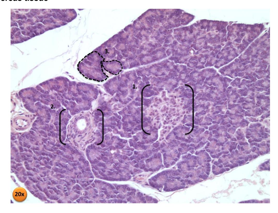
Figure 15.28 Photomicrograph of Pancreas Section.
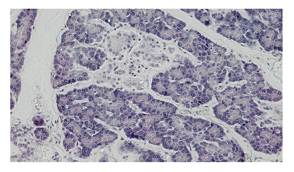
Figure 15.29 Endocrine and Exocrine Pancreas. Cross Section: Human Pancreas Magnification: 200x, Hematoxylin Eosin Stain.
15.22 Post Lab Activities
15.23 Test Your Understanding
15.24 Post Lab Activity 15.1
15.25 Labeling
Label the organs of the endocrine system.
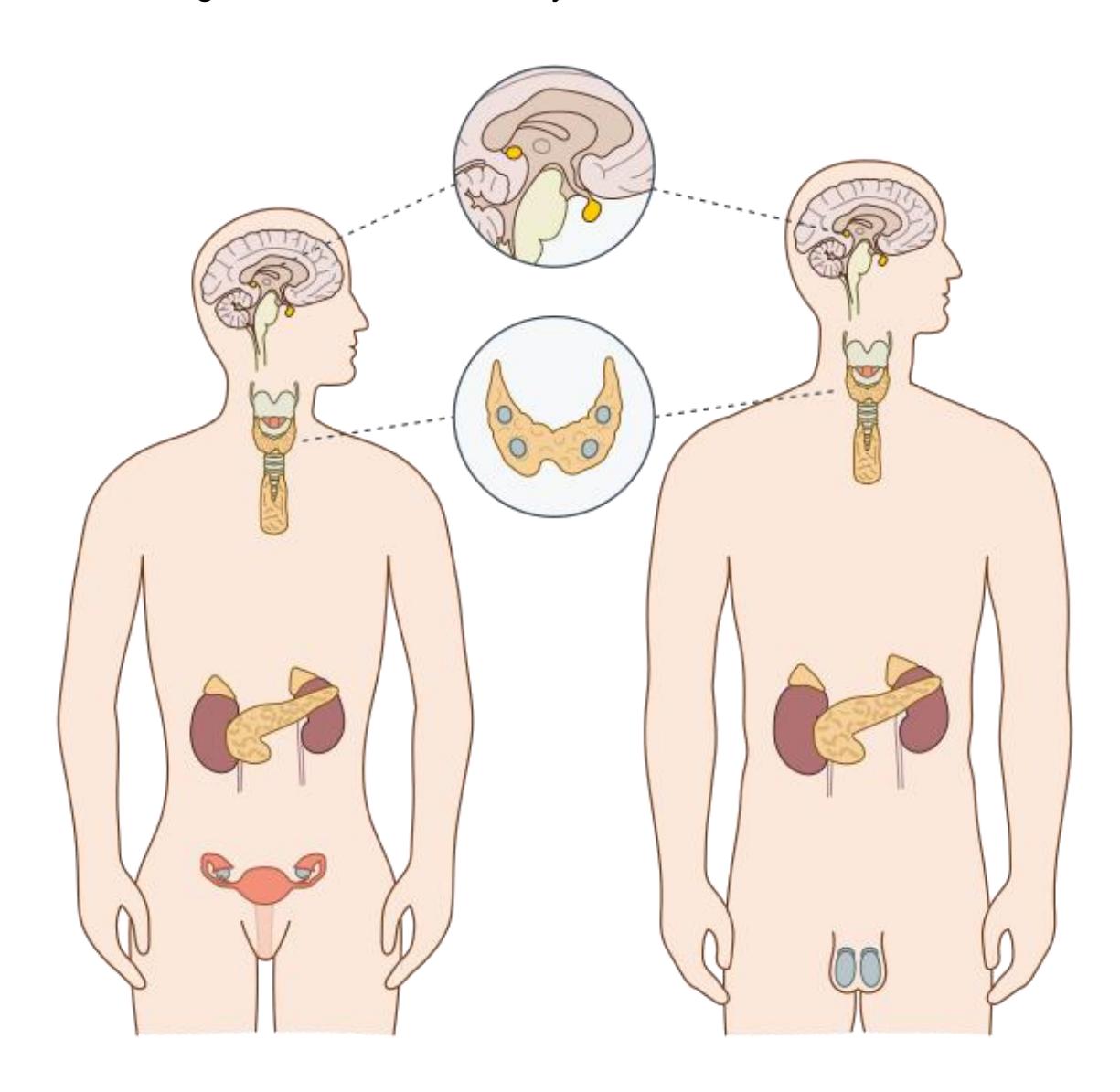
Figure 15.30 Unlabeled Endocrine System.
15.26 Post Lab Activity 15.2
15.27 Identification
Identify the structures in the thyroid.
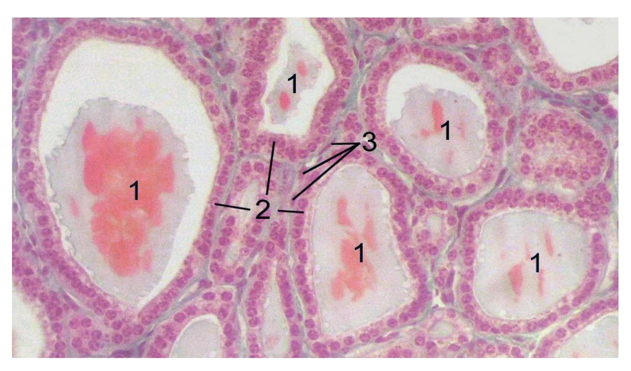
Figure 15.31. Section of a thyroid gland under the microscope.
- ______________________________________________________________
- ______________________________________________________________
- ______________________________________________________________
15.28 Post Lab Activity 15.3
15.29 Crossword Puzzles
Complete the crossword puzzles below.
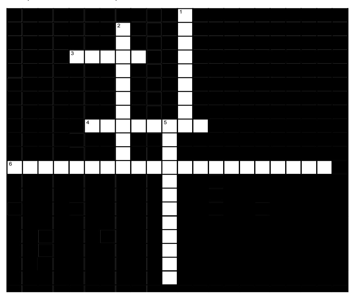
Figure 15.32 Crossword of the Pituitary.
15.29.1 Across
3 Circulating hormones are released into the _____ by an endocrine gland and must travel to reach their (often distant) target. (5)
4 _____ enter the cell and interact directly with DNA and nuclear factors to bring about changes in gene expression. (8)
6 Together, the ____ and _____ regulate the secretion of virtually every hormone in the body. (12,9)
15.29.2 Down
1 _____ based hormones are made from an amino acid by chemical modification or made by joining amino acids to form a peptide (very smallx protein). (5,4)
2 Steroids, one of the two major hormone classes, are made from _____. (11)
5 Hormones are transported from the hypothalamus to the pituitary via the _____. (12)
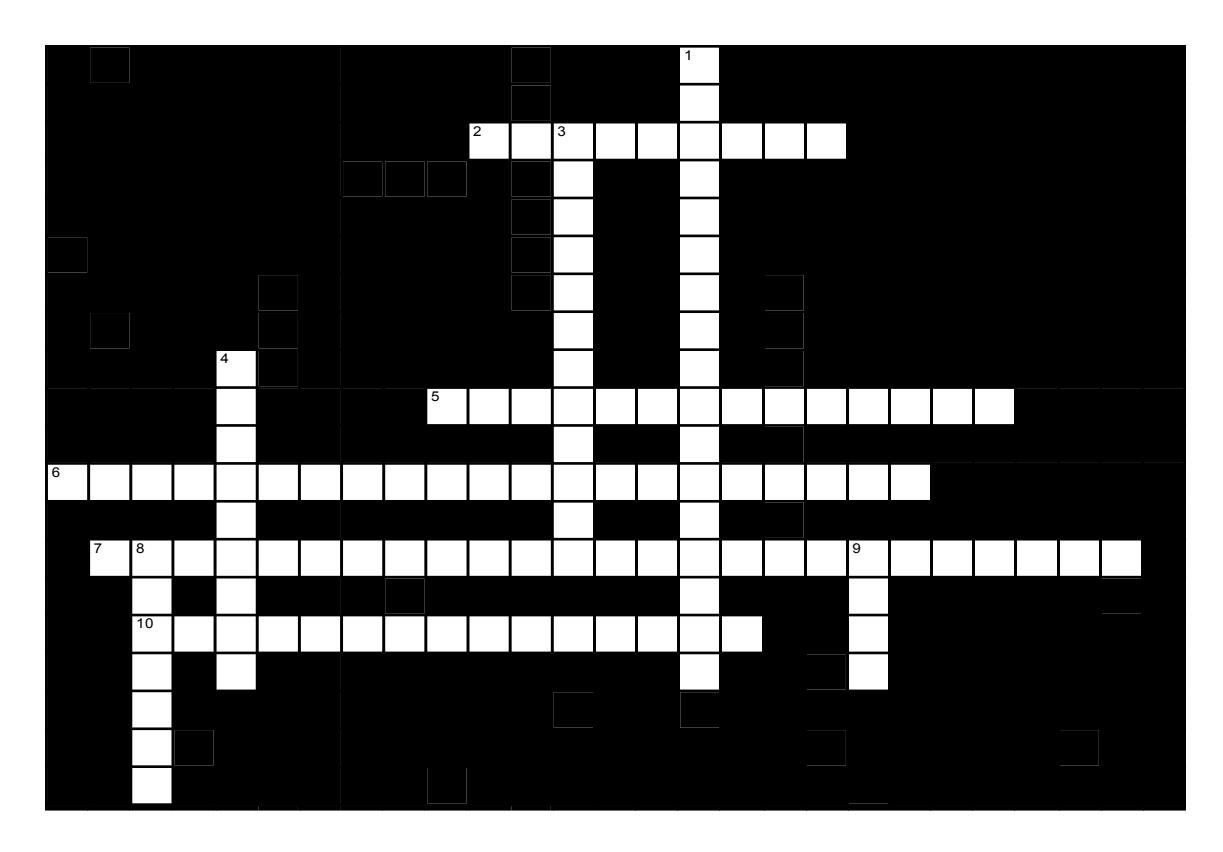
Figure 15.33 Crossword of the Endocrine Hormones.
15.29.3 Across
- 2 In the pancreas, _____ cells produce insulin; _____ cells produce glucagon. (4,5)
- 5 Epinephrine and norepinephrine are released into the blood by the _____. (7,7)
- 6 Antidiuretic hormone (ADH) promotes _____ by the ____. (5,9,7)
- 7 _____ stimulates milk production by the breasts and is produced by the _____. (8,8,9)
- 10 the anterior portion of the pituitary is the _____ (15)
15.29.4 Down
- 1 The third hormone produced by the thyroid is unlike the other two: it helps to regulate _____ metabolism and is called _____. (7,10)
- 3 In the male, _____ initiates maturation of male reproductive organs, causes appearance of secondary sexual characteristics and sex drive, is necessary for sperm production, and maintains sex organs in their functional state. (12)
- 4 PTH _____ calcium in the blood. (9)
- 8 In addition to their role in pregnancy, hormones produced by the _____ are responsible for maturation of the reproductive organs, breast development, and the menstrual cycle. (This question, of course, refers to females (7)
- 9 The phrase ‘thyroid hormone’ refers actually to two hormones, _____ and _____. (2,2)
15.30 Answer Keys
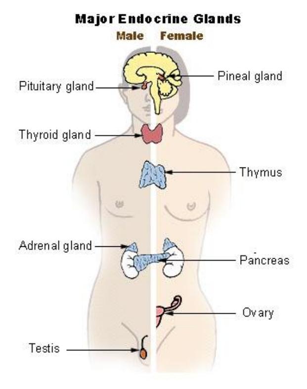
Figure 15.34 Labeled Endocrine System.
15.30.1 Crosswords
15.31 Figure 15.32. Crossword of the pituitary
- Across: 3 Blood, 4 Steroids, 6 Hypothalamus pituitary.
- Down: 1 Amino acid, 2 Cholesterol, 5 Infundibulum.
15.31.1 Figure 15.33. Crossword of the endocrine hormones.
- Across: 2 Beta alpha, 5 Adrenal medulla, 6 Water retention kidneys, 7 Prolactin, anterior pituitary, 10 Adenohypophysis.
- Down: 1 Calcium calcitonin, 3 Testosterone, 4 Increases, 8 Ovaries, 9 T3 T4.
Chapter 15: Endocrine System
| Key Terms | Definitions |
|---|---|
| acetylcholine (ACh) | neurotransmitter that binds at a motor endplate to trigger depolarization |
| adrenal medulla | interior portion of the adrenal (or suprarenal) gland that releases epinephrine and norepinephrine into the bloodstream as hormones |
| adrenergic | synapse where norepinephrine is released, which binds to α- or β- adrenergic receptors |
| afferent branch | component of a reflex arc that represents the input from a sensory neuron, for either a special or general sense |
| agonist | any exogenous substance that binds to a receptor and produces a similar effect to the endogenous ligand |
| alpha (α)-adrenergic receptor | one of the receptors to which epinephrine and norepinephrine bind, which comes in two subtypes: α1 and α2 |
| antagonist | any exogenous substance that binds to a receptor and produces an opposing effect to the endogenous ligand |
| anticholinergic drugs | drugs that interrupt or reduce the function of the parasympathetic system |
| baroreceptor | mechanoreceptor that senses the stretch of blood vessels to indicate changes in blood pressure |
| beta (β)-adrenergic receptor | one of the receptors to which epinephrine and norepinephrine bind, which comes in three subtypes: β1, β2, and β3 |
| cardiac accelerator nerves | preganglionic sympathetic fibers that cause the heart rate to increase when the cardiovascular center in the medulla initiates a signal |
| cardiovascular center | region in the medulla that controls the cardiovascular system through cardiac accelerator nerves and vasomotor nerves, which are components of the sympathetic division of the autonomic nervous system |
| celiac ganglion | one of the collateral ganglia of the sympathetic system that projects to the digestive system |
| central neuron | specifically referring to the cell body of a neuron in the autonomic system that is located in the central nervous system, specifically the lateral horn of the spinal cord or a brain stem nucleus |
| cholinergic synapse | synapse at which acetylcholine is released and binds to the nicotinic or muscarinic receptor |
| ciliary ganglion | one of the terminal ganglia of the parasympathetic system, located in the posterior orbit, axons from which project to the iris |
| craniosacral system | alternate name for the parasympathetic division of the autonomic nervous system that is based on the anatomical location of central neurons in brain-stem nuclei and the lateral horn of the sacral spinal cord; also referred to as craniosacral outflow |
| efferent branch | component of a reflex arc that represents the output, with the target being an effector, such as muscle or glandular tissue |
| endogenous | describes substance made in the human body |
| endogenous chemical | substance produced and released within the body to interact with a receptor protein |
| epinephrine | signaling molecule released from the adrenal medulla into the bloodstream as part of the sympathetic response |
| exogenous | describes substance made outside of the human body |
| exogenous chemical | substance from a source outside the body, whether it be another organism such as a plant or from the synthetic processes of a laboratory, which binds to a transmembrane receptor protein |
| fight-or-flight response | set of responses induced by sympathetic activity that lead to either fleeing a threat or standing up to it, which in the modern world is often associated with anxious feelings |
| ganglionic neuron | specifically refers to the cell body of a neuron in the autonomic system that is located in a ganglion |
| gray rami communicantes (singular = ramus communicans) | unmyelinated structures that provide a short connection from a sympathetic chain ganglion to the spinal nerve that contains the postganglionic sympathetic fiber |
| intramural ganglia | terminal ganglia of the parasympathetic system that are found within the walls of the target effector |
| ligand-gated cation channel | ion channel, such as the nicotinic receptor, which is specific to positively charged ions and opens when a molecule such as a neurotransmitter binds to it |
| limbic lobe | structures arranged around the edges of the cerebrum that are involved in memory and emotion |
| long reflex | reflex arc that includes the central nervous system |
| muscarinic receptor | type of acetylcholine receptor protein that is characterized by also binding to muscarine and is a metabotropic receptor |
| nicotinic receptor | type of acetylcholine receptor protein that is characterized by also binding to nicotine and is an ionotropic receptor |
| norepinephrine | signaling molecule released as a neurotransmitter by most postganglionic sympathetic fibers as part of the sympathetic response, or as a hormone into the bloodstream from the adrenal medulla |
| parasympathetic division | division of the autonomic nervous system responsible for restful and digestive functions |
| parasympathomimetic drugs | drugs that enhance or mimic the function of the parasympathetic system |
| paravertebral ganglia | autonomic ganglia superior to the sympathetic chain ganglia |
| postganglionic fiber | axon from a ganglionic neuron in the autonomic nervous system that projects to and synapses with the target effector; sometimes referred to as a postganglionic neuron |
| preganglionic fiber | axon from a central neuron in the autonomic nervous system that projects to and synapses with a ganglionic neuron; sometimes referred to as a preganglionic neuron |
| prevertebral ganglia | autonomic ganglia that are anterior to the vertebral column and functionally related to the sympathetic chain ganglia |
| reflex arc | circuit of a reflex that involves a sensory input and motor output, or an afferent branch and an efferent branch, and an integrating center to connect the two branches |
| rest and digest | set of functions associated with the parasympathetic system that lead to restful actions and digestion |
| short reflex | reflex arc that does not include any components of the central nervous system |
| somatic reflex | reflex involving skeletal muscle as the effector, under the control of the somatic nervous system |
| sympathetic chain ganglia | series of ganglia adjacent to the vertebral column that receive input from central sympathetic neurons |
| sympathetic division | division of the autonomic nervous system associated with the fight-or-flight response |
| sympatholytic drug | drug that interrupts, or ‘lyses,’ the function of the sympathetic system |
| target effector | organ, tissue, or gland that will respond to the control of an autonomic or somatic or endocrine signal |
| terminal ganglia | ganglia of the parasympathetic division of the autonomic system, which are located near or within the target effector, the latter also known as intramural ganglia |
| thoracolumbar system | alternate name for the sympathetic division of the autonomic nervous system that is based on the anatomical location of central neurons in the lateral horn of the thoracic and upper lumbar spinal cord |
| vasomotor nerves | preganglionic sympathetic fibers that cause the constriction of blood vessels in response to signals from the cardiovascular center |
| visceral reflex | reflex involving an internal organ as the effector, under the control of the autonomic nervous system |
| white rami communicantes (singular = ramus communicans) | myelinated structures that provide a short connection from a sympathetic chain ganglion to the spinal nerve that contains the preganglionic sympathetic fiber |
| adrenal cortex | outer region of the adrenal glands consisting of multiple layers of epithelial cells and capillary networks that produces mineralocorticoids and glucocorticoids |
| adrenal glands | endocrine glands located at the top of each kidney that are important for the regulation of the stress response, blood pressure and blood volume, water homeostasis, and electrolyte levels |
| adrenal medulla | inner layer of the adrenal glands that plays an important role in the stress response by producing epinephrine and norepinephrine |
| adrenocorticotropic hormone (ACTH) anterior pituitary | hormone that stimulates the adrenal cortex to secrete corticosteroid hormones (also called corticotropin) |
| alarm reaction | the short-term stress, or the fight-or flight response, of stage one of the general adaptation syndrome mediated by the hormone’s epinephrine and norepinephrine |
| aldosterone | hormone produced and secreted by the adrenal cortex that stimulates sodium and fluid retention and increases blood volume and blood pressure |
| alpha cell | pancreatic islet cell type that produces the hormone glucagon |
| Anterior pituitary | The pituitary gland consists of three lobes: anterior pituitary, pars intermedia, and posterior pituitary. The anterior pituitary releases hormones that regulate growth, reproduction, lactation, and stress. |
| angiotensin- converting enzyme | the enzyme that converts angiotensin I to angiotensin II |
| antidiuretic hormone (ADH) | hypothalamic hormone that is stored by the posterior pituitary and that signals the kidneys to reabsorb water |
| atrial natriuretic peptide (ANP) | peptide hormone produced by the walls of the atria in response to high blood pressure, blood volume, or blood sodium that reduces the reabsorption of sodium and water in the kidneys and promotes vasodilation |
| autocrine | chemical signal that elicits a response in the same cell that secreted it |
| beta cell | pancreatic islet cell type that produces the hormone insulin |
| calcitonin peptide | hormone produced and secreted by the parafollicular cells (C cells) of the thyroid gland that functions to decrease blood calcium levels |
| colloid | viscous fluid in the central cavity of thyroid follicles, containing the glycoprotein thyroglobulin |
| cortisol | glucocorticoid important in gluconeogenesis, the catabolism of glycogen, and downregulation of the immune system |
| cyclic adenosine monophosphate (cAMP) | second messenger that, in response to adenylyl cyclase activation, triggers a phosphorylation cascade |
| delta cell | minor cell type in the pancreas that secretes the hormone somatostatin |
| diabetes mellitus | condition caused by destruction or dysfunction of the beta cells of the pancreas or cellular resistance to insulin that results in abnormally high blood glucose levels |
| endocrine gland | tissue or organ that secretes hormones into the blood and lymph without ducts such that they may be transported to organs distant from the site of secretion |
| endocrine system | cells, tissues, and organs that secrete hormones as a primary or secondary function and play an integral role in normal bodily processes |
| epinephrine | primary and most potent catecholamine hormone secreted by the adrenal medulla in response to short-term stress; also called adrenaline |
| erythropoietin (EPO) | protein hormone secreted in response to low oxygen levels that triggers the bone marrow to produce red blood cells |
| estrogens | class of predominantly female sex hormones important for the development and growth of the female reproductive tract, secondary sex characteristics, the female reproductive cycle, and the maintenance of pregnancy |
| exocrine system | cells, tissues, and organs that secrete substances directly to target tissues via glandular ducts |
| first messenger | f hormone that binds to a cell membrane hormone receptor and triggers activation of a second messenger system |
| follicle-stimulating hormone (FSH) | anterior pituitary hormone that stimulates the production and maturation of sex cells |
| G protein | protein associated with a cell membrane hormone receptor that initiates the next step in a second messenger system upon activation by hormone- receptor binding |
| general adaptation syndrome (GAS) | human body’s three-stage response pattern to short- and long-term stress |
| gigantism | disorder in children caused when abnormally high levels of GH prompt excessive growth |
| glucagon | pancreatic hormone that stimulates the catabolism of glycogen to glucose, thereby increasing blood glucose levels |
| glucocorticoids | hormones produced by the zona fasciculata of the adrenal cortex that influence glucose metabolism |
| goiter | enlargement of the thyroid gland either as a result of iodine deficiency or hyperthyroidism |
| gonadotropins | hormones that regulate the function of the gonads |
| growth hormone (GH) | anterior pituitary hormone that promotes tissue building and influences nutrient metabolism (also called somatotropin) |
| hormone | secretion of an endocrine organ that travels via the bloodstream or lymphatics to induce a response in target cells or tissues in another part of the body |
| hormone receptor | protein within a cell or on the cell membrane that binds a hormone, initiating the target cell response |
| hyperglycemia | abnormally high blood glucose levels |
| hyperparathyroidism | disorder caused by overproduction of PTH that results in abnormally elevated blood calcium |
| hyperthyroidism clinically abnormal, elevated level of | clinically abnormal, elevated level of thyroid hormone in the blood; characterized by an increased metabolic rate, excess body heat, sweating, diarrhea, weight loss, and increased heart rate |
| hypoparathyroidism | disorder caused by underproduction of PTH that results in abnormally low blood calcium |
| hypophyseal portal system | network of blood vessels that enables hypothalamic hormones to travel into the anterior lobe of the pituitary without entering the systemic circulation |
| hypothalamus | region of the diencephalon inferior to the thalamus that functions in neural and endocrine signaling |
| hypothyroidism | clinically abnormal, low level of thyroid hormone in the blood; characterized by low metabolic rate, weight gain, cold extremities, constipation, and reduced mental activity |
| infundibulum | stalk containing vasculature and neural tissue that connects the pituitary gland to the hypothalamus (also called the pituitary stalk) |
| inhibin | hormone secreted by the male and female gonads that inhibits FSH production by the anterior pituitary |
| insulin | pancreatic hormone that enhances the cellular uptake and utilization of glucose, thereby decreasing blood glucose levels |
| leptin | protein hormone secreted by adipose tissues in response to food consumption that promotes satiety |
| luteinizing hormone (LH) | anterior pituitary hormone that triggers ovulation and the production of ovarian hormones, and the production of testosterone |
| melatonin | amino acid-derived hormone that is secreted in response to low light and causes drowsiness |
| mineralocorticoids | hormones produced by the zona glomerulosa cells of the adrenal cortex that influence fluid and electrolyte balance |
| neonatal hypothyroidism | condition characterized by cognitive deficits, short stature, and other signs and symptoms in people born to people who were iodine-deficient during pregnancy |
| norepinephrine | secondary catecholamine hormone secreted by the adrenal medulla in response to short-term stress; also called noradrenaline |
| osmoreceptor | hypothalamic sensory receptor that is stimulated by changes in solute concentration (osmotic pressure) in the blood |
| ovary | Ovaries are found on either side of the uterus. They produce eggs and hormones for menstruation and pregnancy. |
| oxytocin | hypothalamic hormone stored in the posterior pituitary gland and important in stimulating uterine contractions in labor, milk ejection during breastfeeding, and feelings of attachment (produced by males and females) |
| pancreas | organ with both exocrine and endocrine functions located posterior to the stomach that is important for digestion and the regulation of blood glucose |
| pancreatic islets | specialized clusters of pancreatic cells that have endocrine functions; also called islets of Langerhans |
| paracrine | chemical signal that elicits a response in neighboring cells; also called paracrine factor |
| parathyroid glands | small, round glands embedded in the posterior thyroid gland that produce parathyroid hormone (PTH) |
| parathyroid hormone (PTH) | ) peptide hormone produced and secreted by the parathyroid glands in response to low blood calcium levels |
| pineal gland | endocrine gland that secretes melatonin, which is important in regulating the sleep-wake cycle |
| pituitary dwarfism | disorder in children caused when abnormally low levels of GH result in growth retardation |
| pituitary gland | bean-sized organ suspended from the hypothalamus that produces, stores, and secretes hormones in response to hypothalamic stimulation (also called hypophysis) |
| Posterior pituitary | The posterior pituitary, an extension of the hypothalamus, produces and release oxytocin and antidiuretic hormone (ADH). |
| progesterone | predominantly female sex hormone important in regulating the female reproductive cycle and the maintenance of pregnancy |
| prolactin (PRL) | anterior pituitary hormone that promotes development of the mammary glands and the production of breast milk |
| second messenger | molecule that initiates a signaling cascade in response to hormone binding on a cell membrane receptor and activation of a G protein |
| Testicle (testis) | A testicle or testis is the gonad in all male bilaterians, including humans, and is homologous to the ovary in females. Its primary functions are the production of sperm and the secretion of androgens, primarily testosterone. |
| testosterone | steroid hormone secreted by the testes and important in the maturation of sperm cells, growth and development of the reproductive system, and the development of secondary sex characteristics |
| thymosins | hormones produced and secreted by the thymus that play an important role in the development and differentiation of T cells |
| thymus | organ that is involved in the development and maturation of T-cells and is particularly active during infancy and childhood |
| thyroid gland thyroid-stimulating hormone (TSH) | large endocrine gland responsible for the synthesis of thyroid hormones anterior pituitary hormone that triggers secretion of thyroid hormones by the thyroid gland (also called thyrotropin) |
| thyroxine (also, tetraiodothyronine, T4) | amino acid-derived thyroid hormone that is more abundant but less potent than T3 and often converted to T3 by target cells |
| triiodothyronine (also, T3) | amino acid-derived thyroid hormone that is less abundant but more potent than T4 |
| zona fasciculata | intermediate region of the adrenal cortex that produce hormones called glucocorticoids |
| zona glomerulosa | most superficial region of the adrenal cortex, which produces the hormones collectively referred to as mineralocorticoids |
| zona reticularis | deepest region of the adrenal cortex, which produces the steroid sex hormones called androgens |