10 Skeletal Muscle Histology and Microanatomy
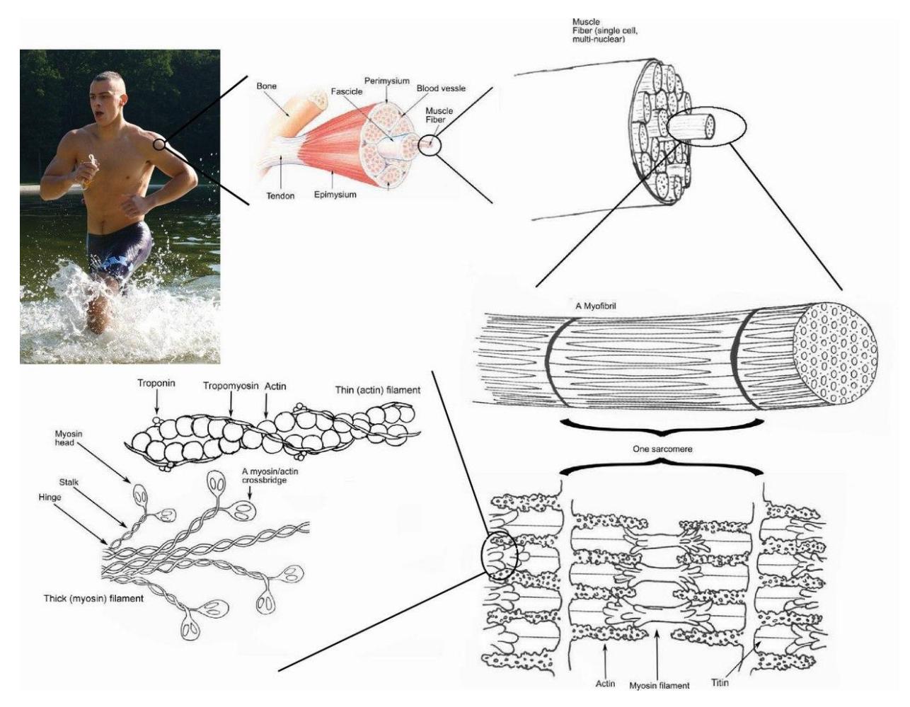
Figure 10.1 Skeletal Muscle Histology
10.1 Objectives
The objectives of this lab are to complete the following:
10.2 Muscle Tissue:
- Identify where you can find each muscle tissue in the body.
- Compare and contrast the three muscle tissue types.
- Skeletal muscle
- Long cord-like, nonbranching
- Voluntary
- Striations
- Multiple Nuclei
- Smooth muscle
- Tapered
- Involuntary
- Single nucleus
- Not striated
- Cardiac muscle
- branching
- Striations
- Intercalated discs
- Single Nucleus
In a model or diagram of the muscle fiber, describe the locations and functions of the following structures:
Endomysium
Sarcolemma
T-tubule
Mitochondria
Sarcoplasmic reticulum
Sarcomere
Myofibril
Myofibril
Neuromuscular junction (NMJ)
In a model or diagram of skeletal muscle (cross-section), describe the locations and functions of the following:
Epimysium
Perimysium
Fascicle
Endomysium
Muscle fiber
In a model or diagram of skeletal muscle myofibril, label and describe the locations and functions of the following:
- Myofilaments
- Z disc (line)
- M line
- I band.
- H zone
- A band
- Thick filament
- Myosin
- Thin filament
- Actin
- Troponin
- Tropomyosin
In a model or diagram of the neuromuscular junction, describe the structure and functions of the neuromuscular junction:
Axon
Axon terminals
Synaptic vesicles
Synaptic cleft
Synaptic vesicles
Acetylcholine
Motor neuron
Axon terminal
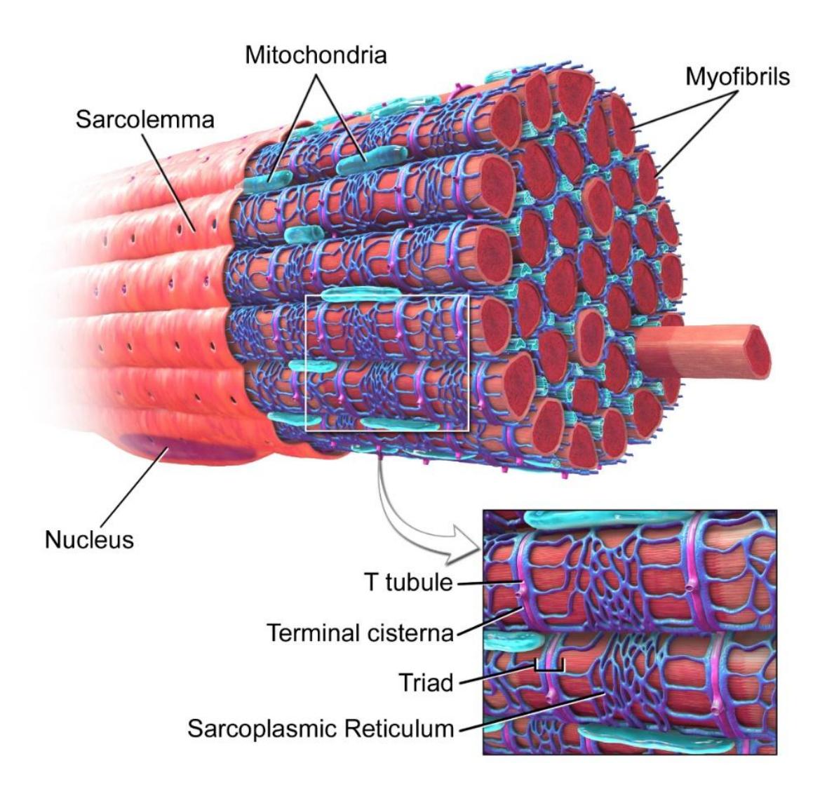
Figure 10.2 Skeletal Muscle Fiber.
10.3 Prelab Activities
10.4 Prelab Activity 10.1
10.5 Definitions Muscle Structures
| Term | Definition |
|---|---|
| tendon | |
| epimysium | |
| fascicle | |
| muscle fiber | |
| endomysium | |
| sarcolemma | |
| nucleus | |
| sarcoplasm | |
| mitochondria | |
| sarcoplasmic reticulum | |
| T or transverse tubules | |
| myofibrils | |
| myofilaments | |
| actin | |
| tropomyosin | |
| Thin filament | |
| Thick filament | |
| Myofibrils | |
| Myofibril Structure | |
| sarcomere | |
| Z discs | |
| M line | |
| I band | |
| A band (dark) | |
| H zone |
10.6 Prelab Activity 10.2
10.7 Labeling
Label the structures, membranes, or layers of the muscle in figures 10.3 and 10.4.
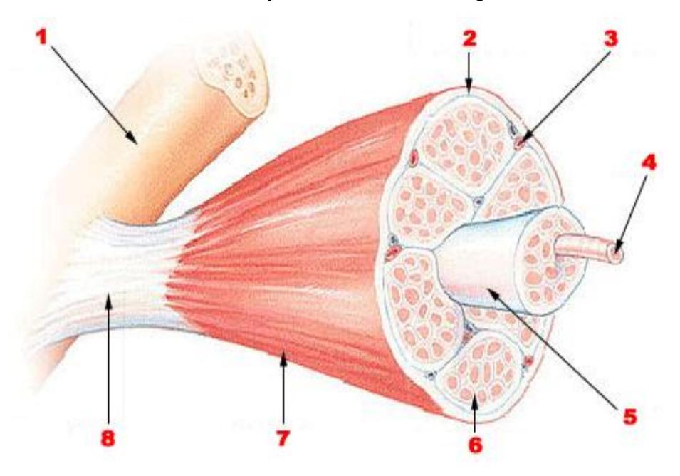
Figure 10.3 Unlabeled Skeletal Muscle.
Table 10.1 Structure Identification
| Structure Number | Structure Identification |
|---|---|
| 1 | Organ: |
| 2 | Connective tissue layer: |
| 3 | Organelle: |
| 4 | Structure name: |
| 5 | Bundle: |
| 6 | Connective tissue layer: |
| 7 | Connective tissue layer: |
| 8 | Structure: |
10.7.1 Label the following structures of the skeletal muscle:
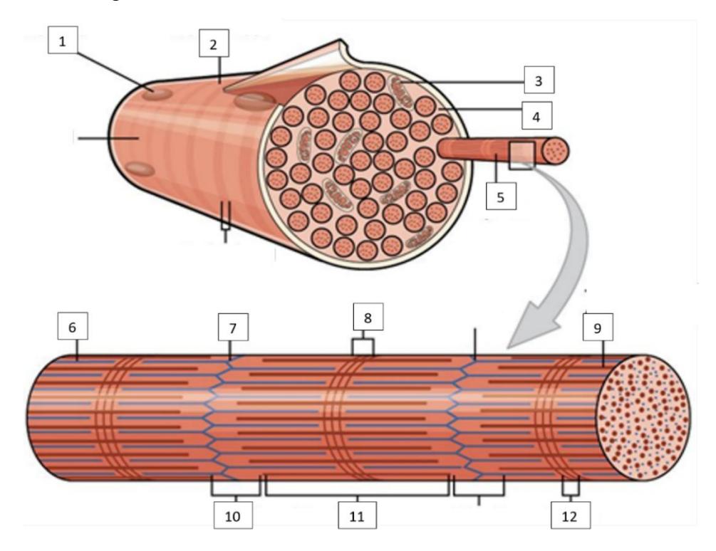
Figure 10.4 Unlabeled Muscle Fiber.
Table 10.2 Muscle Histology Identification
| Structure Number | Structure Identification |
|---|---|
| 1 | Organelle: |
| 2 | Surface layer: |
| 3 | Organelle: |
| 4 | Internal gel: |
| 5 | Filament name: |
| 6 | Filament type: |
| 7 | line: |
| 8 | Band: |
| 9 | Filament type: |
| 10 | Band: |
| 11 | Band: |
| 12 | Band: |
10.8 Lab Activities
10.9 Skeletal Muscle Cell Microscopic Structure and Gross Structures
10.10 Muscle Tissue Types
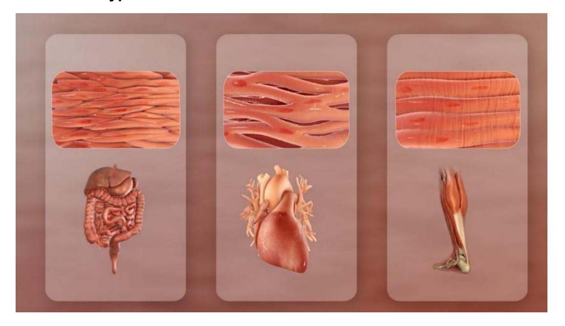
Figure 10.5 Three Muscle Types (L to R: Smooth, Cardiac, and Skeletal Muscles).
10.11 Lab Activity 10.1
Compare and contrast the three muscle tissue types: Skeletal muscle, Smooth muscle, and Cardiac muscle.
- Working in groups of two get a microscope from the storage cabinet and a slide of the skeletal, cardiac and muscle cells.
- Focus on the slide and once you have found the images on the slides provided in the lab, increase your magnification to 400x. Find a section where you can clearly identify the unique structural features described in the background information of your textbook. Draw and label the microscopic structures of all each of the three muscle tissue types in the spaces provided.
- Next take a picture with your cellphone. Upload the screenshot into a PPT folder and label the parts of each muscle tissue.
- Draw and label the three types of muscle cells in the table below.
| Cell type: | Cell type: |
|---|---|
| Magnification: | Magnification: |
| Cell type: | Cell type: |
|---|---|
| Magnification: | Magnification: |
The following cells may be used for review after class. Virtual muscle review source from University of Michigan Virtual Microscope:
https://histology.medicine.umich.edu/
- Skeletal muscle, longitudinal section, H&E, 83X: https://histologyslides.med.umich.edu/Histology/Basic%20Tissues/Muscle/05 8thin\_HISTO\_83X.htm
- Heart, ventricle, H&E, 40X (cardiac muscle, intercalated discs). See Cardiovascular section. https://histologyslides.med.umich.edu/Histology/Cardiovascular%20System/0 98-1\_HISTO\_40X.htm
- Smooth muscle, Esophagus and stomach, H&E, 40X https://histologyslides.med.umich.edu/Histology/Digestive%20System/Pharyn x%20Esophagus%20and%20Stomach/155\_HISTO\_40X.htm
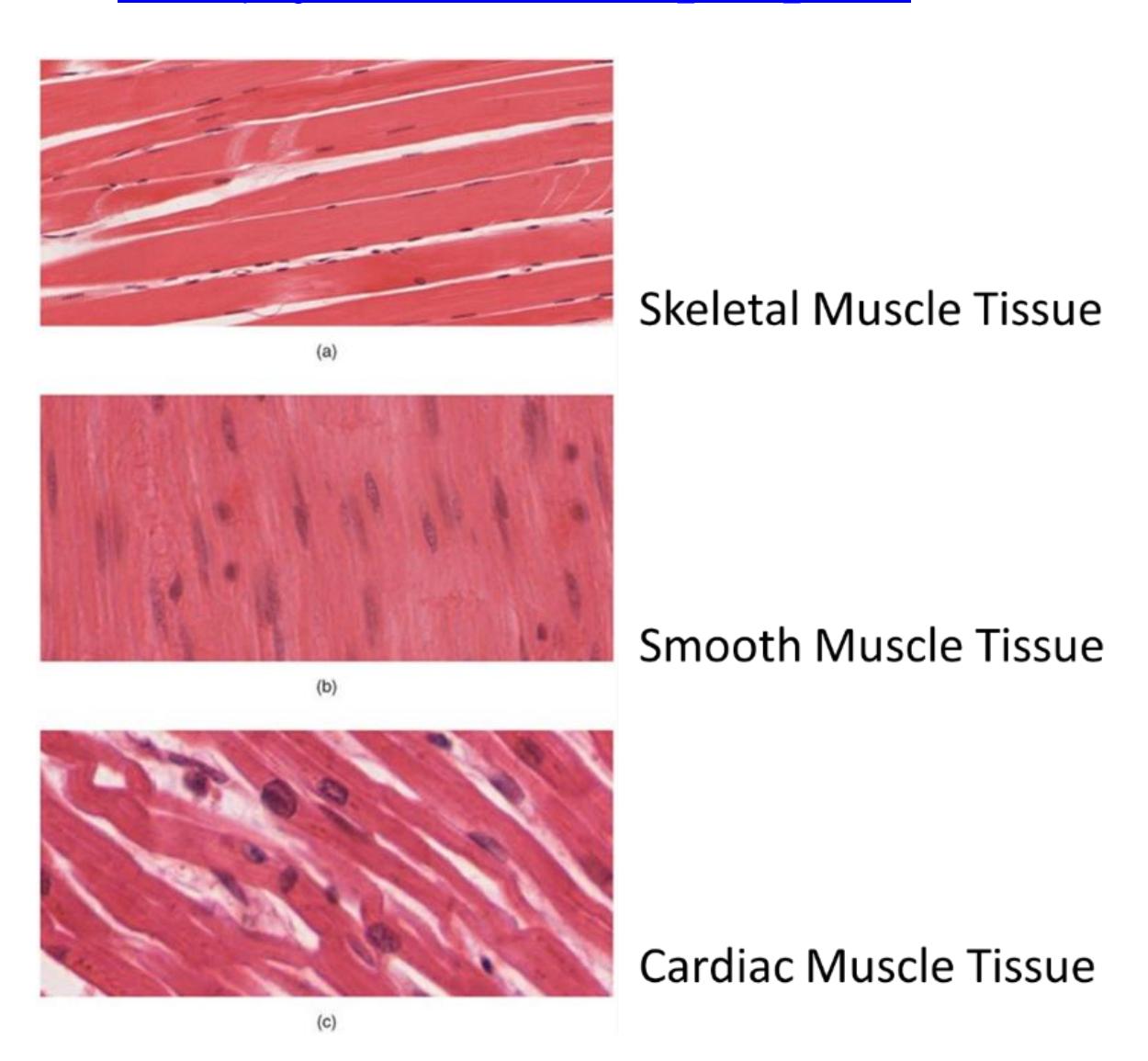
Figure 10.6 Three Types of Muscle Fibers.
10.12 Gross Anatomy of a Muscle
10.13 Lab Activity 10.2
Identify the following terms on a diagram or model.
- Go to the area where the models are kept and obtain the model of the microanatomy human muscle fiber.
- Collaborate with your group to identify and label the structures found in the regions of the outer, middle, and inner ear.
- Invite the group next to you to check your work, then have your instructor check your work.
- Take a picture of the work for later reference before the upcoming practical and if you have time have a classmate record a video while you review the structures of the ear you need to review.
10.14 Identify the Following Muscle Structures
- Muscle layers
- Muscle fiber
- Fascicle
- Myofibril
- Connective tissue layers
- Epimysium
- Perimysium
- Endomysium
- Other structures
- Tendon
- Aponeurosis
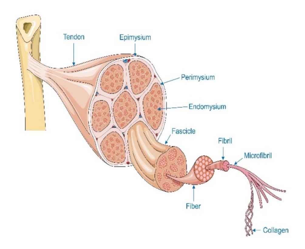
Figure 10.7 Connective Tissue Layers in a Muscle.
10.15 Muscle Fiber Organization
10.16 Lab Activity 10.3
- Using the model of the microanatomy human muscle fiber.
- Locate and label the muscle features listed below.
- Invite the group next to you to review your model identifications for correctness.
- Finally take pictures of the model toad to your study guide.
10.17 Sarcomere Structures in the Myofibril
10.17.1 Terms
Sarcolemma Sarcoplasm Sarcoplasmic reticulum T tubules Nuclei Mitochondria Myofibril
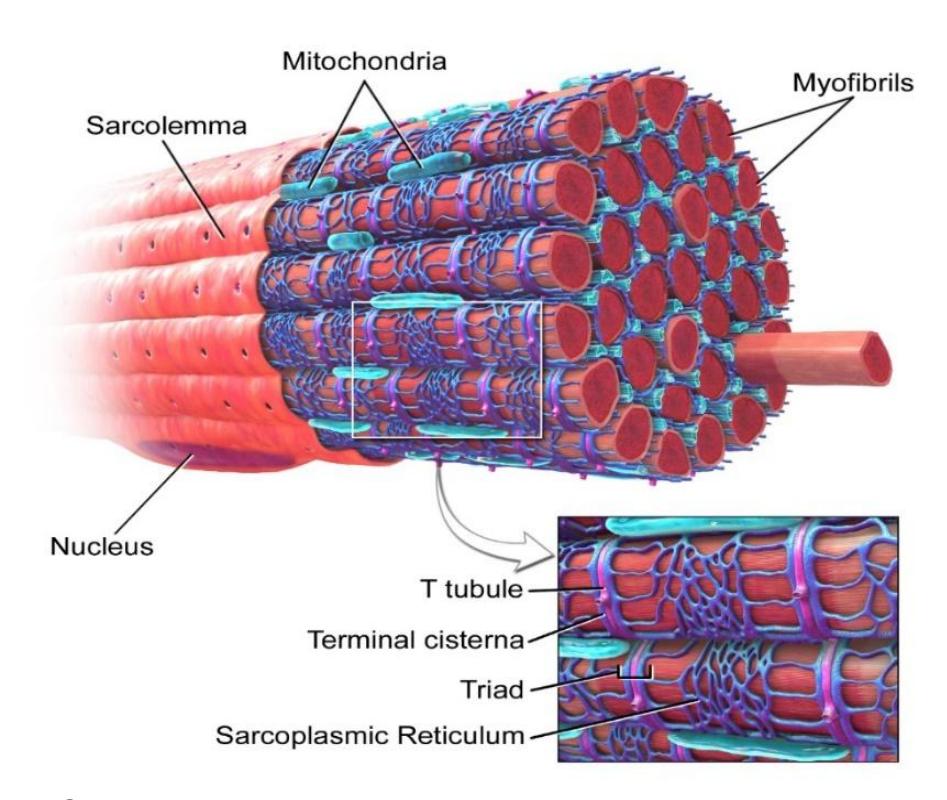
Figure 10.8 Skeletal Muscle Fiber.
10.18 Lab Activity 10.4:
Obtain the model of the sarcomere from your storage area for this exercise.
Identify and label the components of the sarcomere in the list below.
Using your cellphone create a five-minute video explaining the structure and function of the sarcomere.
Sarcomere
A band
I band.
Z disc (Z line)
H zone
M line
Thick filament
Myosin
Thin filament
Actin
Troponin
Tropomyosin
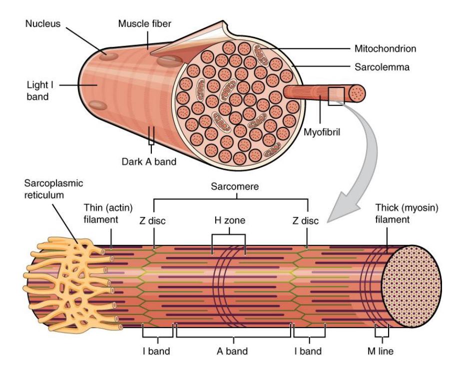
Figure 10.9 Organization of a Muscle Fiber.
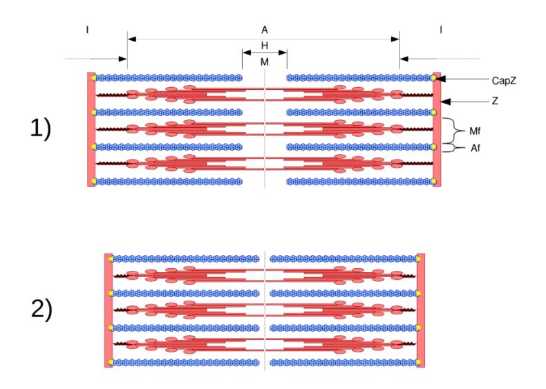
Figure 10.10 Sarcomere Structure in the 1) Relaxed and 2) Contracted States
10.19 The Neuromuscular Junction
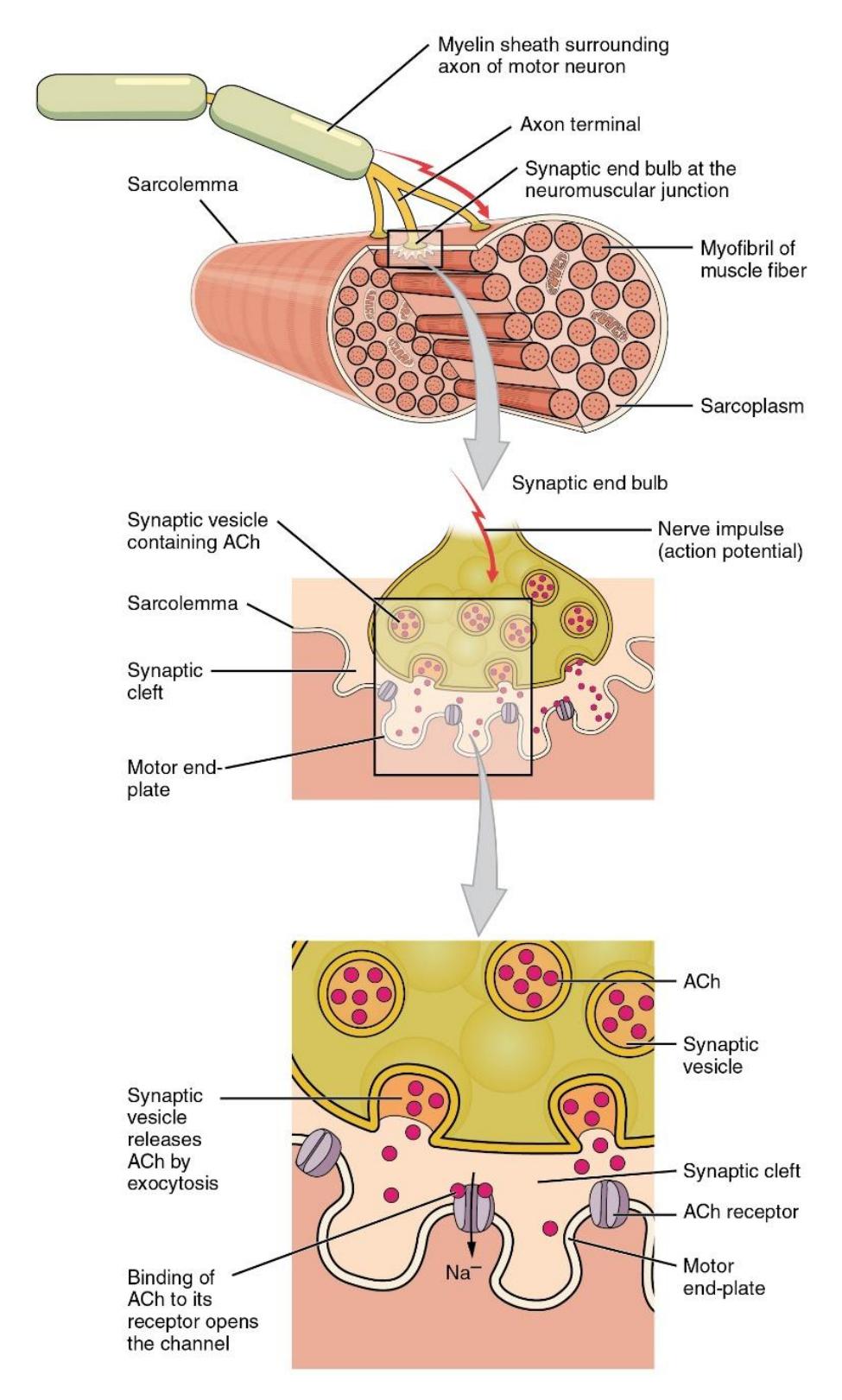
Figure 10.11 The Neuromuscular Junction.
10.20 Review Videos
- Muscle microanatomy:https://youtu.be/E9cNTG7oe88
- Visible Body | The Structure of Skeletal Muscle: https://youtu.be/TUD\_wlAB5Z0
- Anatomy of a muscle cell; Khan Academy https://youtu.be/uY2ZOsCnXIA
- Skeletal Muscle Microanatomy Anatomy BIO2113: https://youtu.be/YhUVOkGpgJs
10.20.1 Review Materials
Virtual Muscle Material Source: University of Michigan Virtual Microscope: https://histology.medicine.umich.edu/
- Skeletal muscle, longitudinal section, H&E, 83X: https://histologyslides.med.umich.edu/Histology/Basic%20Tissues/Muscle/05 8thin\_HISTO\_83X.htm
- Heart, ventricle, H&E, 40X (cardiac muscle, intercalated discs). See Cardiovascular section. https://histologyslides.med.umich.edu/Histology/Cardiovascular%20System/0 98-1\_HISTO\_40X.htm
- Smooth muscle, Esophagus and stomach, H&E, 40X https://histologyslides.med.umich.edu/Histology/Digestive%20System/Pharyn x%20Esophagus%20and%20Stomach/155\_HISTO\_40X.htm
10.21 Post Lab Activities
10.22 Check Your Understanding
10.23 Post Lab Activity 10.1
10.24 Matching
Table 10.1 Identify the Muscle Tissue Type (Note: there may be more than one correct muscle type per answer)
| Answers | Characteristics | Choices |
|---|---|---|
| Involuntary | Cardiac | |
| Banded/striated | Smooth | |
| Coordinated activity – acts in a pump | Skeletal | |
| Moves facial skin and the bones | ||
| Voluntary | ||
| Tapered | ||
| Longitudinal and circular layers | ||
| Cord-like | ||
| Multiple nuclei | ||
| Branched | ||
| Single nucleus |
10.25 Table 10.2 Identify the Facia Type
Write the correct choice or choices in the “Answers” column.
| Answers | Definitions | Choices |
|---|---|---|
| Muscle fiber cell membrane | Endomysium | |
| Surrounds the whole muscle | Perimysium | |
| Connective tissue that covers the body under the skin as well as the whole muscles. | Exomysium | |
| Thin layer of connective tissue that surrounds the muscle fibers cell membrane | Sarcolemma | |
| Surrounds a group of fasciculi. | Facia |
10.26 Post Lab Activity 10.2
10.26.0.1 Identification
Identify and label the parts of the myofibril.
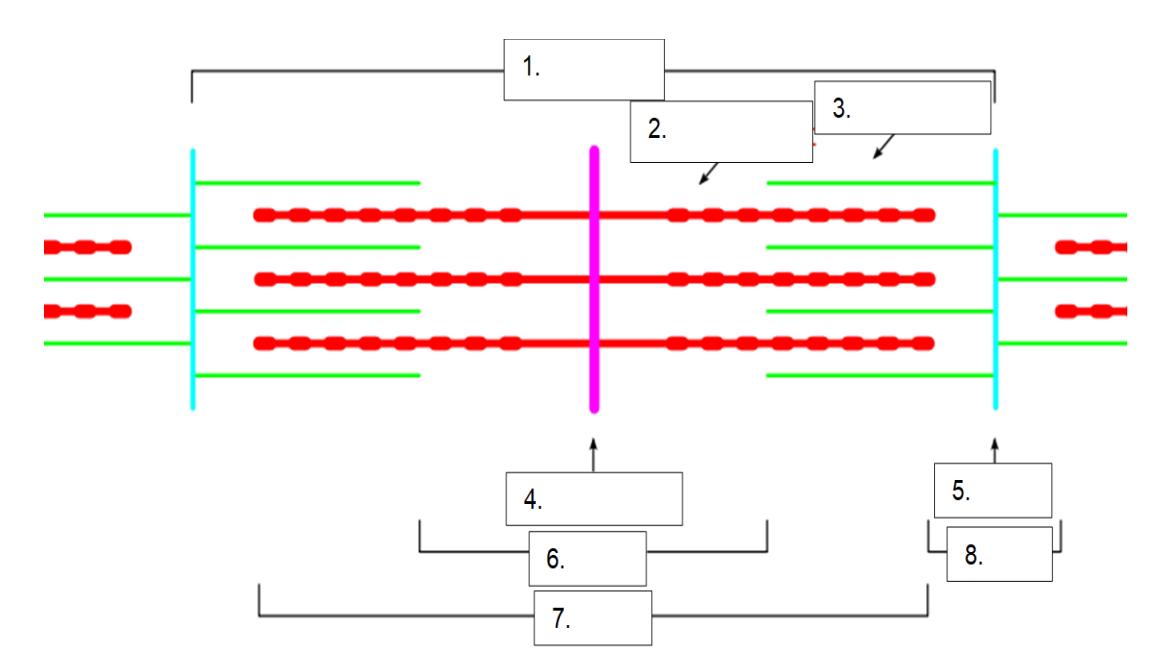
Figure 10.12 Unlabeled Sarcomere.
- ____________________________________
- ____________________________________
- ____________________________________
- ____________________________________
- ____________________________________
- ____________________________________
- ____________________________________
- ____________________________________
10.27 Post Lab Activity 10.3
10.27.1 Crossword Puzzles
Complete the crossword puzzles.
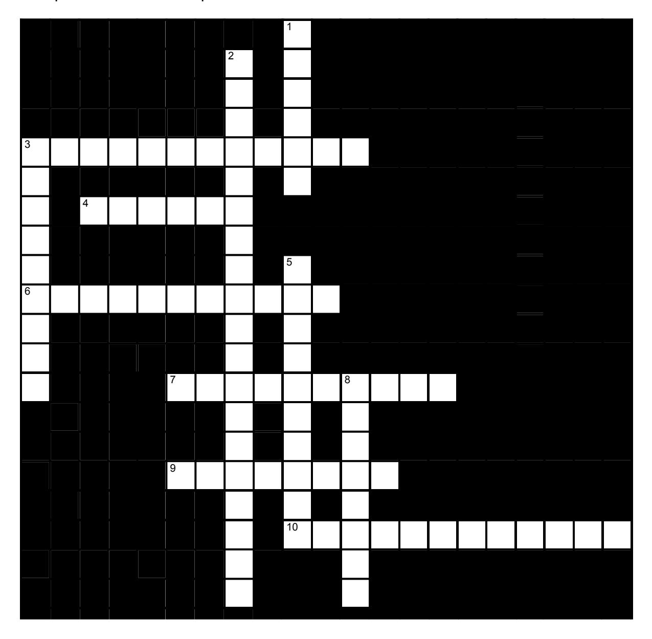
Figure 10.13 Skeletal Muscle Fibers.
10.27.2 Across
- 3 The myofibrils of _____ muscle are not aligned, so that the muscle appears to be _____. (6,6)
- 4 Muscle cells are also called muscle _____. (6)
- 6 There are two types of _____, actin and myosin. (11)
- 7 Within a skeletal muscle, a connective tissue membrane called the ____ surrounds each fascicle (10)
- 9 A group of muscle fibers is called a muscle _____, which means ‘bundle’. (8)
- 10 Cardiac muscle fibers contain large numbers of _____, which provides the power to allow them to have incredible endurance. (12)
10.27.3 Down
- 1 For many muscles, the connective tissue membranes within the muscle merge at the ends with a connective tissue ‘rope’ called a(n) _____. (6)
- 2 When a muscle attaches directly to a bone, rather than to a tendon which is attached to a bone, the _____ of the muscle fuses with the _____ of the bone. (9,10)
- 3 A(n) _____ is the smallest contractile unit of a skeletal muscle: it extends from one Z-disk to the next. (9)
- 5 Each muscle fiber is enclosed in a delicate, areolar connective tissue sheath called the _____. (10)
- 8 Much of the heat in the body is produced by _____ muscle. (8)
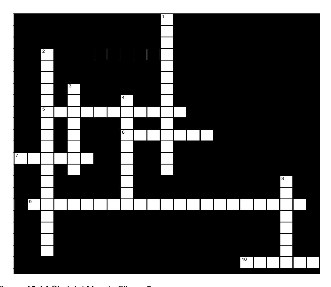
Figure 10.14 Skeletal Muscle Fibers-2.
- 5 There are two types of _____, actin and myosin. (11)
- 6 Action potentials that are carried deep into the cell signal the sarcoplasmic reticulum to _____. (7)
- 7 In a skeletal muscle fiber, after tropomyosin shifts in response to calcium, _____ binds to actin. (6)
- 9 The smooth endoplasmic reticulum (smooth ER) of muscle cells is called the _____. (12,9)
- 10 _____ are found at each end of the sarcomere. (1,5)
10.27.4 Down
8 Skeletal muscle fibers are formed when several stem cells merge: as a result, skeletal muscle fibers have _____ (8,6)
10.28 Crossword Puzzle Solutions
10.29 Figure 10.13. Skeletal muscle fibers.
- Across: 5 Myofilament, 6 Calcium, 7 Myosin, 9 Sarcoplasmic reticulum, 10 Z disks.
- Down: 1 Multiple nuclei, 2 Actin microfilament, 3 Troponin, 4 Sarcolemma, 8 T tubules.
10.30 Figure 10.14. Skeletal muscle fibers 2.
- Across: 3 Smooth smooth, 4 Fibers, 6 Myofilament, 7 Perimysium, 9 Fascicle, 10 Mitochondria.
- Down: 1 Tendon, 2 Epimysium periosteum, 3 Sarcomere, 5 Endomysium, 8 Skeletal.
Chapter 10: Skeletal Muscle Histology and Microanatomy Glossary
| Terms | Definitions |
|---|---|
| acetylcholine (ACh) | neurotransmitter that binds at a motor endplate to trigger depolarization |
| actin | protein that makes up most of the thin myofilaments in a sarcomere muscle fiber |
| action potential | change in voltage of a cell membrane in response to a stimulus that results in transmission of an electrical signal; unique to neurons and muscle fibers |
| aerobic respiration | production of ATP in the presence of oxygen |
| aponeurosis | tendon-like sheet of connective tissue that attaches a skeletal muscle to another skeletal muscle or to a bone |
| ATPase | enzyme that hydrolyzes ATP to ADP |
| atrophy | loss of structural proteins from muscle fibers |
| cardiac muscle | striated muscle found in the heart; joined to one another at intercalated discs and under the regulation of pacemaker cells, which contract as one unit to pump blood through the circulatory system. Cardiac muscle is under involuntary control. |
| concentric contraction | muscle contraction that shortens the muscle to move a load |
| contractility | ability to shorten (contract) forcibly |
| contraction phase twitch | contraction phase when tension increases |
| creatine phosphate | phosphagen used to store energy from ATP and transfer it to muscle |
| depolarize | to reduce the voltage difference between the inside and outside of a cell’s plasma membrane (the sarcolemma for a muscle fiber), making the inside less negative than at rest |
| desmosome | structure that anchors the ends of cardiac muscle fibers to allow contraction to occur |
| eccentric contraction | muscle contraction that lengthens the muscle as the tension is diminished |
| elasticity | ability to stretch and rebound |
| endomysium | loose, and well-hydrated connective tissue covering each muscle fiber in a skeletal muscle |
| epimysium | outer layer of connective tissue around a skeletal muscle |
| excitability | ability to undergo neural stimulation |
| excitation-contraction coupling | sequence of events from motor neuron signaling to a skeletal muscle fiber to contraction of the fiber’s sarcomeres |
| extensibility | ability to lengthen (extend) |
| fascicle | bundle of muscle fibers within a skeletal muscle |
| fast glycolytic (FG) muscle | fiber that primarily uses anaerobic glycolysis |
| fast oxidative (FO) intermediate muscle | fiber that is between slow oxidative and fast glycolytic fibers |
| fibrosis | replacement of muscle fibers by scar tissue |
| glycolysis | anaerobic breakdown of glucose to ATP |
| H zone | The H zone is the bright region of the A band in a sarcomere that only has thick filaments. |
| hyperplasia | process in which one cell splits to produce new cells |
| hypertrophy | addition of structural proteins to muscle fibers |
| hypotonia | abnormally low muscle tone caused by the absence of low-level contractions |
| I band | I band is the light band of the sarcomere in skeletal muscle tissue that contains actin filaments. |
| intercalated disc | part of the sarcolemma that connects cardiac tissue, and contains gap junctions and desmosomes |
| isometric contraction | muscle contraction that occurs with no change in muscle length |
| isotonic contraction | muscle contraction that involves changes in muscle length |
| lactic acid | product of anaerobic glycolysis |
| latent period | the time when a twitch does not produce contraction |
| M line | the center of the sarcomere that runs through the middle of the myosin filaments |
| motor endplate | sarcolemma of muscle fiber at the neuromuscular junction, with receptors for the neurotransmitter acetylcholine |
| motor unit | motor neuron and the group of muscle fibers it innervates |
| Muscle fiber | the individual cells that make up muscle tissue, specifically skeletal muscle, which is responsible for voluntary movements in the body. |
| muscle tension | force generated by the contraction of the muscle; tension generated during isotonic contractions and isometric contractions |
| muscle tone | low levels of muscle contraction that occur when a muscle is not producing movement |
| myoblast | muscle-forming stem cell |
| myofibril | long, cylindrical organelle that runs parallel within the muscle fiber and contains the sarcomeres |
| myosin | protein that makes up most of the thick cylindrical myofilament within a sarcomere muscle fiber |
| neuromuscular junction (NMJ) | synapse between the axon terminal of a motor neuron and the section of the membrane of a muscle fiber with receptors for the acetylcholine released by the terminal |
| neurotransmitter signaling | chemical released by nerve terminals that bind to and activate receptors on target cells |
| oxygen debt | amount of oxygen needed to compensate for ATP produced without oxygen during muscle contraction |
| perimysium | connective tissue that bundles skeletal muscle fibers into fascicles within a skeletal muscle |
| power stroke | action of myosin pulling actin inward(toward theM line) |
| pyruvic acid | product of glycolysis that can be used in aerobic respiration or converted to lactic acid |
| recruitment | increase in the number of motor units involved in contraction |
| relaxation phase | period after twitch contraction when tension decreases |
| sarcolemma | plasma membrane of a skeletal muscle fiber |
| sarcomere | longitudinally, repeating functional unit of skeletal muscle, with all of the contractile and associated proteins involved in contraction |
| sarcoplasm | cytoplasm of a muscle cell |
| sarcoplasmic reticulum (SR) | specialized smooth endoplasmic reticulum, which stores, releases, and retrieves Ca++ |
| satellite cell stem | cell that helps to repair muscle cells |
| skeletal muscle | striated, multinucleated muscle that requires signaling from the nervous system to trigger contraction; most skeletal muscles are referred to as voluntary muscles that move bones and produce movement |
| slow oxidative (SO) muscle | fiber that primarily uses aerobic respiration |
| smooth muscle | nonstriated, mononucleated muscle in the skin that is associated with hair follicles; assists in moving materials in the walls of internal organs, blood vessels, and internal passageways |
| synaptic cleft | space between a nerve (axon) terminal and a motor endplate |
| T-tubule | projection of the sarcolemma into the interior of the cell |
| tendon | a tough band of connective tissue that connects muscles to bones |
| tetanus | a continuous fused contraction |
| thick filament | thick filament the thick myosin strands and their multiple heads projecting from the center of the sarcomere toward, but not all to way to, the Z-discs |
| thin filament | thin filament thin strands of actin and its troponin-tropomyosin complex projecting from the Z-discs toward the center of the sarcomere |
| treppe | stepwise increase in contraction tension |
| triad | the grouping of one T-tubule and two terminal cisternae |
| tropomyosin | regulatory protein that covers myosin binding sites to prevent actin from binding to myosin |
| troponin | regulatory protein that binds to actin, tropomyosin, and calcium |
| twitch | single contraction produced by one action potential |
| visceral muscle | smooth muscle found in the walls of visceral organs |
| voltage-gated sodium channels | proteins that open sodium channels in response to a sufficient voltage change, and initiate and transmit the action potential as Na+ enters through the channel |
| wave summation | addition of successive neural stimuli to produce greater contraction |
| Z disc (z line) | a boundary within the sarcomere, marking the ends of each unit. |