5 Integumentary System
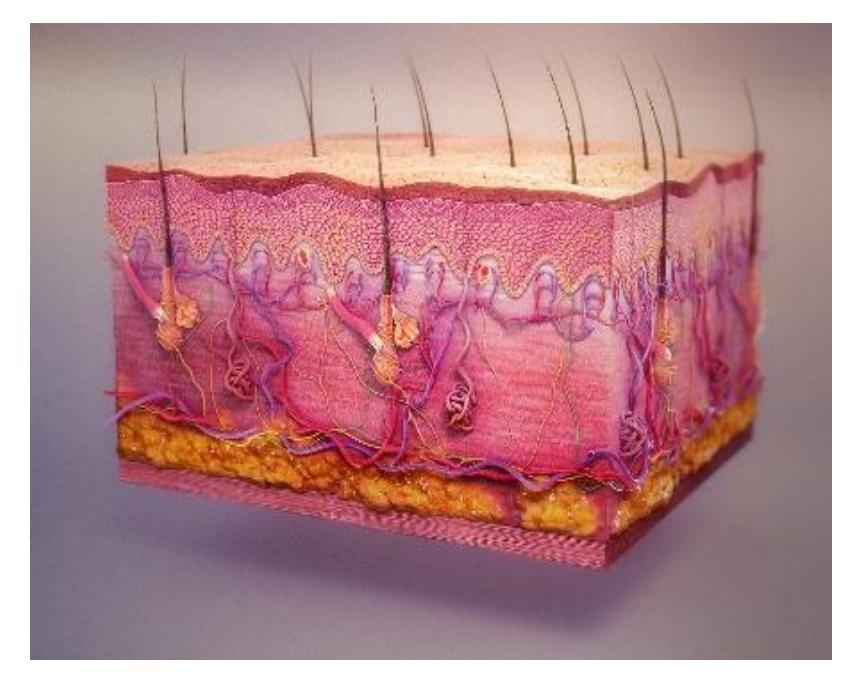
Figure 5.1 The Integumentary System.
5.1 Course Objectives
At the conclusion of this lab, you should be able to:
- Describe the structure of the skin.
- Describe the functions of the integumentary system.
- Differentiate between the layers of the epidermis.
- Identify the components of the dermis and their function.
- Identify the layers of the Integumentary system on a model or a diagram.
- Layers and Sublayers of Cutaneous Membrane also be able to identify the epidermal layers on a model or a diagram (note: the layers are listed from superficial to deep)
- Stratum corneum
- Stratum lucidum thick skin only
- Stratum granulosum
- Stratum spinosum
- Stratum basale (germinitivum)
- Dermis
- Papillary layer
- Reticular layer
- Subcutaneous layer (hypodermis)
- Layers and Sublayers of Cutaneous Membrane also be able to identify the epidermal layers on a model or a diagram (note: the layers are listed from superficial to deep)
- Using a model or diagram identify and describe the function of the following integumentary structures:
- Dermal papillae
- Hair shaft
- Hair root
- Hair follicle
- Pore
- Arrector pili muscle
- Sebaceous gland
- Merocrine (eccrine) sweat gland.
- Apocrine sweat gland
- Free nerve ending
- Meissner’s (tactile) corpuscle
- Pacinian (pressure) corpuscle
- Keratinocyte
- Melanocyte
5.2 Prelab Activities
5.3 Prelab Activity 5.1
Using your textbook define or identify the following terms.
Epidermal Layer
| Term | Definition |
|---|---|
| Epidermis | |
| Stratum corneum | |
| Stratum lucidum | |
| Stratum granulosum | |
| Stratum spinosum | |
| Stratum basale (germinitivum) | |
| Dermal Layer |
Dermal Layer
| Term | Definition |
|---|---|
| Dermis | |
| Papillary layer | |
| Reticular layer |
| Term | Definition |
|---|---|
| Subcutaneous Layer (Hypodermis) |
Integumentary Structures
| Term | Definition |
|---|---|
| Dermal papillae | |
| Hair shaft | |
| Hair root | |
| Hair follicle | |
| Pore | |
| Arrector pili muscle | |
| Sebaceous gland | |
| Merocrine (eccrine) sweat gland | |
| Apocrine sweat gland | |
| Free nerve ending | |
| Meissner’s (tactile) corpuscle | |
| Pacinian (pressure) corpuscle | |
| Keratinocyte | |
| Melanocyte |
5.4 Prelab Activity 5.2
Using your textbook define or identify the following layers.
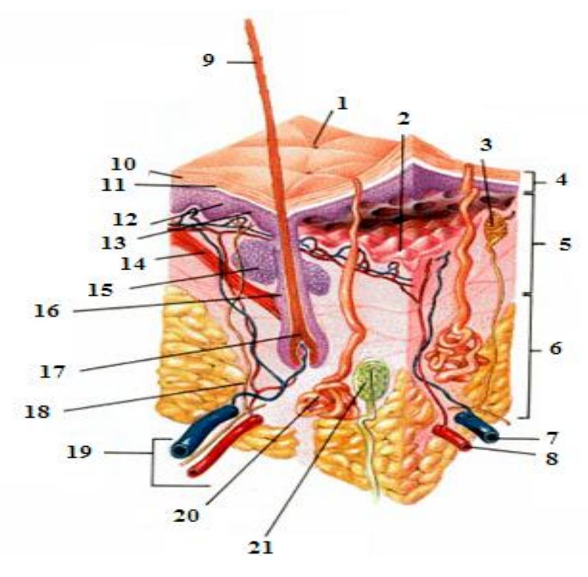
Figure 5.2 The Integumentary System.
5.4.1 Label the following layers.
- 4: ___________________________________
- 5: ___________________________________
- 6: ___________________________________
5.4.2 Label the following accessory structures:
- 15: ___________________________________
- 21: ___________________________________
- 14: ___________________________________
- 20: ___________________________________
5.5 Lab Activities
5.6 Integumentary System
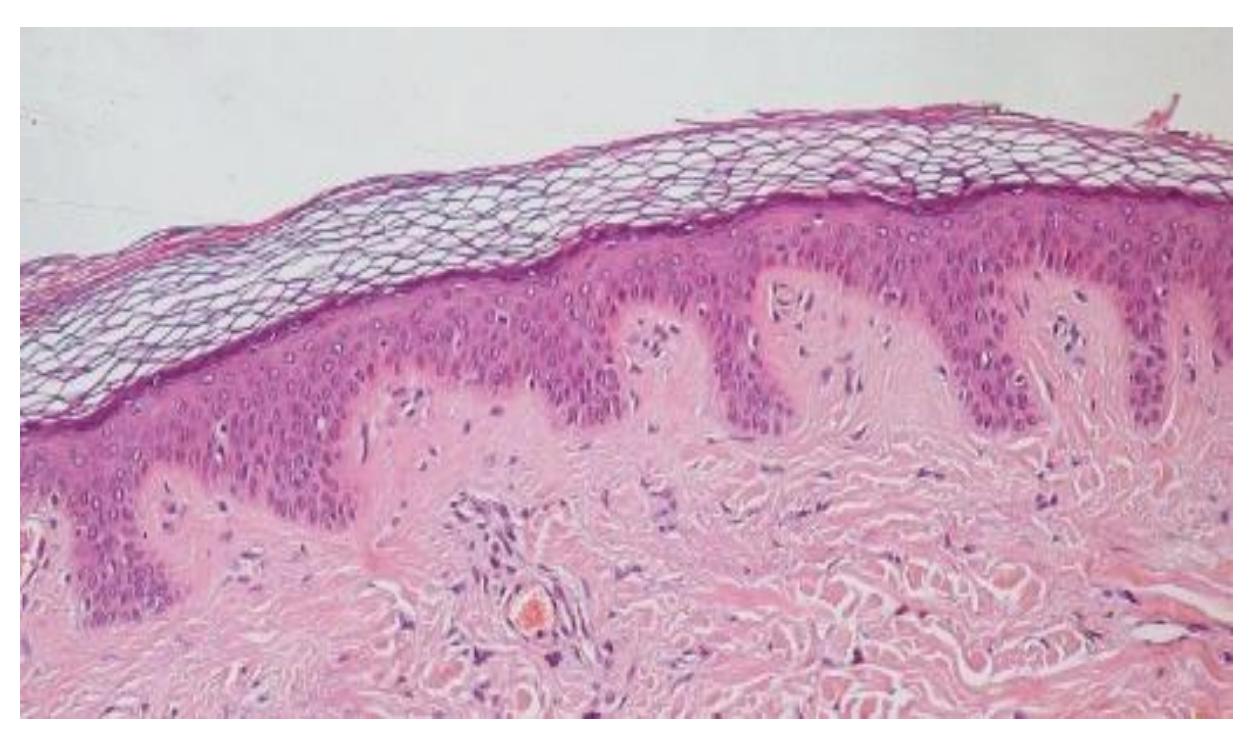
Figure 5.3 Photomicrograph of a Section of Thick Skin.
5.7 General Functions of the Integumentary System
The Integumentary system functions include but are not limited to the following functions:
- protection from physical damage or chemical or biological agents,
- water, and temperature regulation,
- sensory,
- synthesis of vitamin D,
- fat storage.
5.8 Layers of the Skin and the Hypodermis
5.8.1 Terms
- Epidermis
- Dermis
- Hypodermis (Subcutaneous)
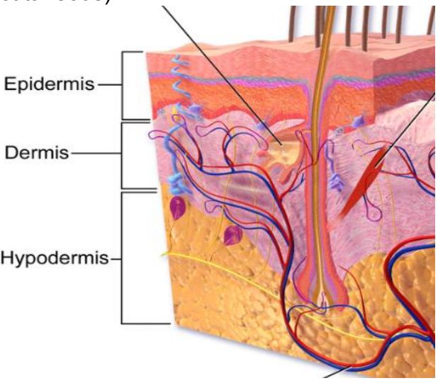
Figure 5.4 Diagram of the Integument Layers.
5.8.2 Epidermal Layers and their Function
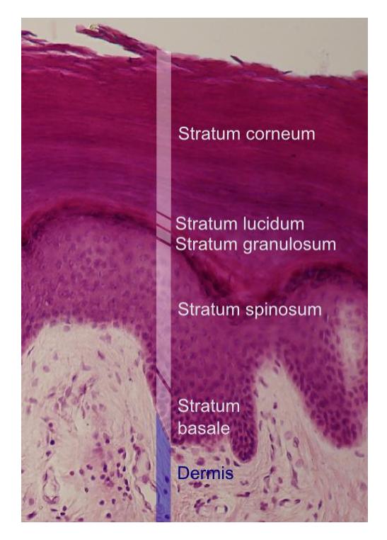
Figure 5.5 Layers of the Epidermis
Table 5.1 Epidermal Layers (Listed Superficial to Deep)
| Epidermal Layer | Major Features |
|---|---|
| Stratum corneum | |
| Stratum lucidum | |
| Stratum granulosum | |
| Stratum spinosum | |
| Stratum basale (germinitivum) |
5.9 Lab Activities
5.10 Lab Activity 5.1
5.11 Anatomy of the Integumentary System
- Obtain a model of Hubbard TM Scientific or a 3D model of the integumentary system.
- Take a picture of one of the models of the integumentary system available in the lab with your cell phone. Label and identify the epidermis, dermis, and the hypodermis.
- Set up a microscope with a slide of the skin. Focus on the integument and take a picture (photomicrograph) of the slide with your cellphone. Identify the epidermis, dermis, and the hypodermis.
- Epidermis.
- Identify the type of cells that comprise this layer.
- Determine if there are blood vessels in this layer.
- Label the apical and basal surfaces of this layer.
- Dermis
- Notice that the interface between the dermal and epidermal layers is uneven. The projections are called dermal papillae. Find this interface on the model before you and label them.
- Do the same for these structures in your picture.
- Epidermis.
5.12 Anatomy of the Epidermis
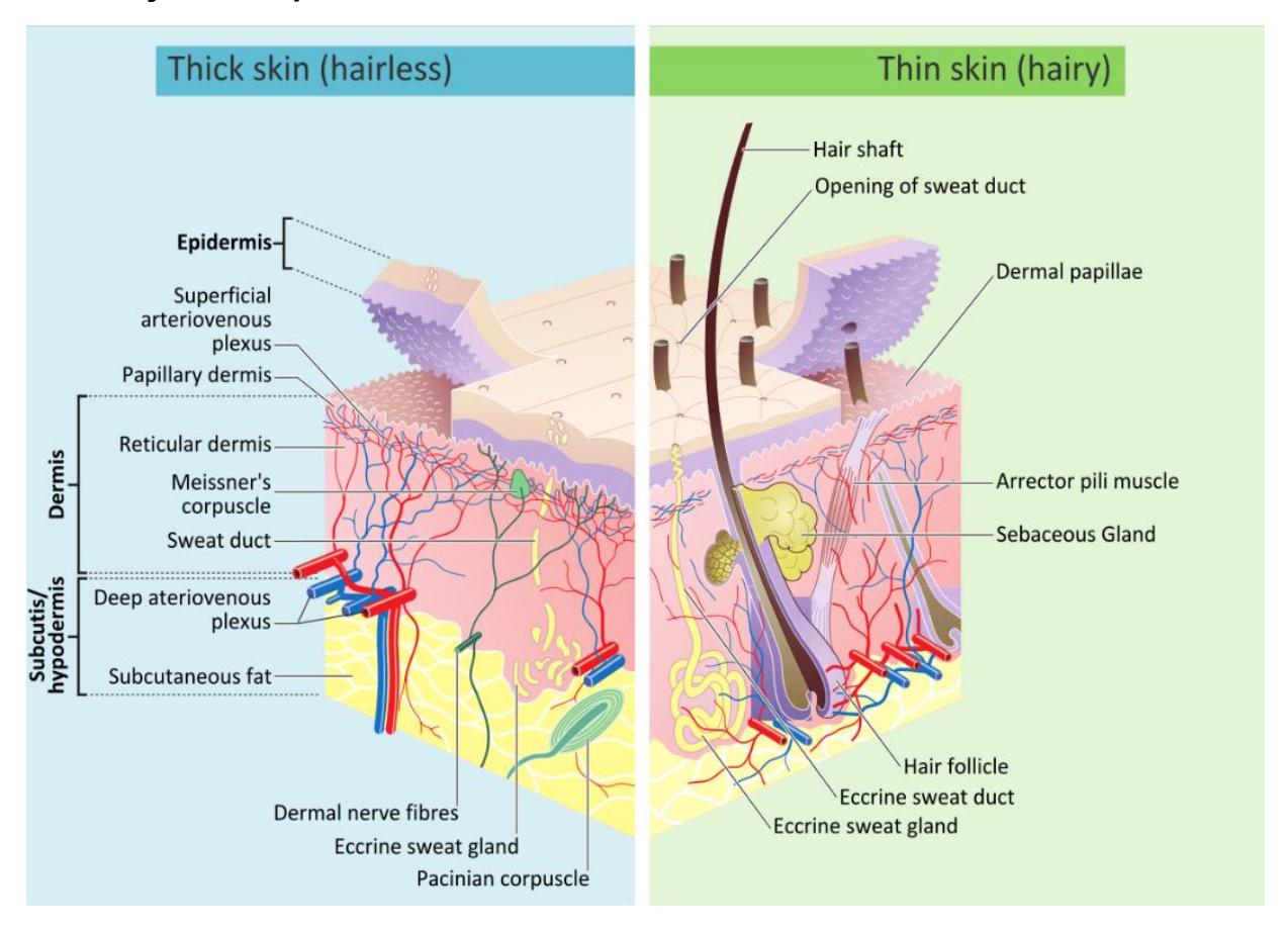
Figure 5.6 Layers of the Integumentary system. Comparison of the structure of thick skin vs thin skin.
5.12.1 Lab Activity 5.1:
5.13 The Anatomy of the Integumentary System continued
- Compare the thick and thin skin models, or pictures, of the skin.
- What differences do you note in their appearance and structure?
- Where is each type located on the body? From this observation provide an explanation of each skin type’s function.
- Identify and describe the layers of each type of skin.
- Find a picture of the epidermis on the internet and print the picture.
- Label all of the layers of the epidermis
- Provide a description of each layer of the epidermis on the table below.
Table 5.2 Epidermal layers listed superficial to deep.
| Epidermal Layer | Description |
|---|---|
| Stratum corneum | |
| Stratum lucidum | |
| Stratum granulosum | |
| Stratum spinosum | |
| Stratum basale |
5.13.1 Lab Activity 5.2:
5.14 Dermis
- Using one of the lab models, with the assistance of the picture below, locate the two layers of the dermis.
- Locate the papillary ridge.
- Identify the type of connective tissue in each layer.
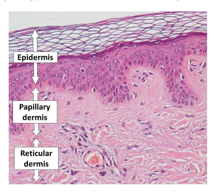
Figure 5.7 Epidermis, Papillary Dermis and Reticular Dermis.
5.15 Lab Activity 5.3:
- Using a model, with the assistance of the following diagram, identify and describe the function of the following integumentary structures.
- Identify and label the structures indicated below.
- Have your instructor check your work.
- Take a picture to use as a study guide.
5.16 Dermal Structures
- Dermal papillae
- Hair shaft
- Hair root
- Hair follicle
- Pore
- Arrector pili muscle
- Sebaceous gland
- Merocrine (eccrine) sweat gland.
- Apocrine sweat gland
- Free nerve ending
- Meissner’s (tactile) corpuscle
- Pacinian (pressure) corpuscle
- Keratinocyte
- Melanocyte
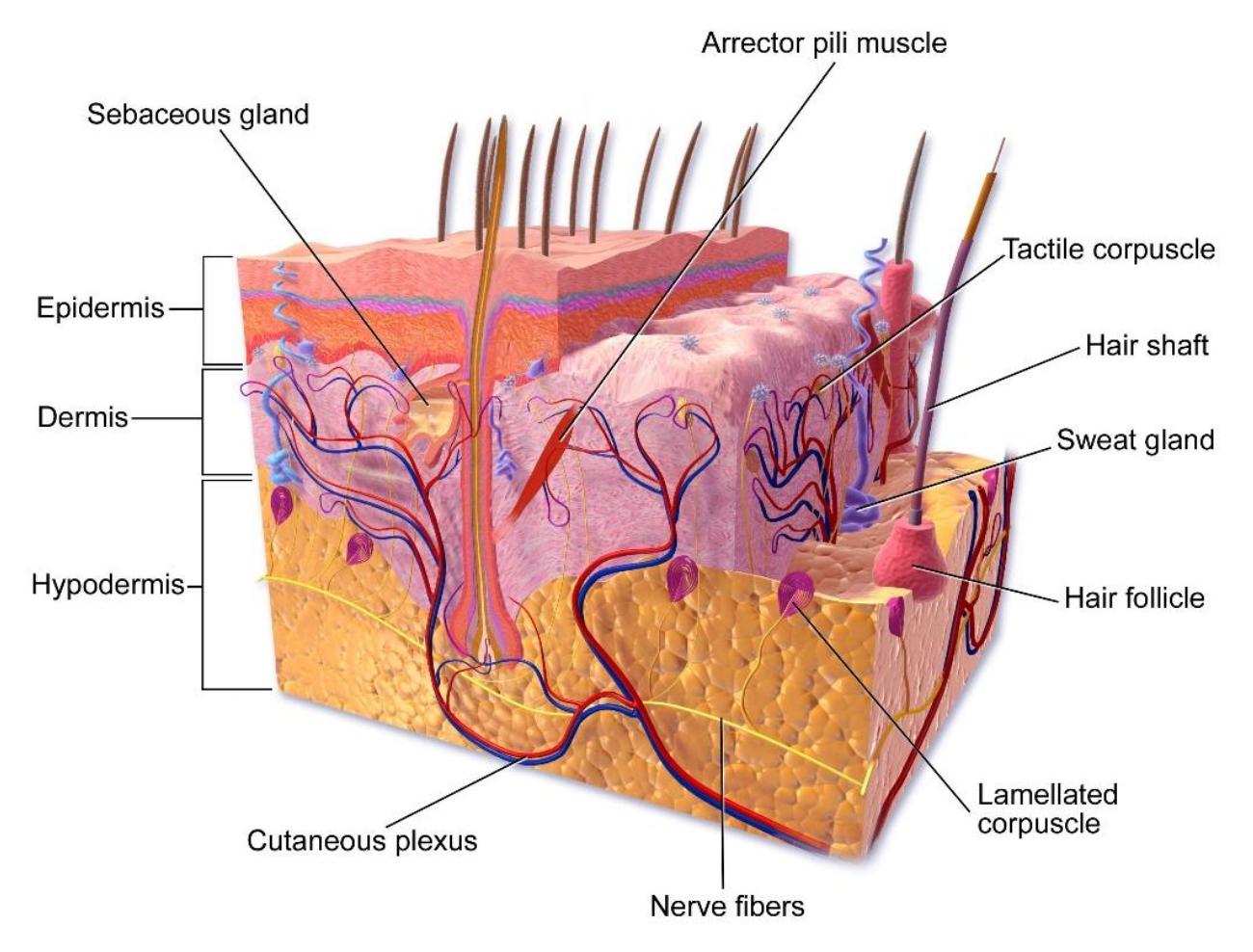
Figure 5.8 Anatomy of Human Skin.
5.17 Lab Activity 5.4:
Describe the function of each of the listed dermal structures.
Table 5.3 Table of the Dermal Structures and their Function
| Dermal Structure | Function(s) |
|---|---|
| Dermal papillae | |
| Pore | |
| Arrector pili muscle | |
| Sebaceous gland | |
| Merocrine (eccrine) sweat gland | |
| Apocrine sweat gland | |
| Free nerve ending | |
| Meissner’s (tactile) corpuscle | |
| Pacinian (pressure) corpuscle | |
| Keratinocyte | |
| Melanocyte |
5.18 Epidermis
The epidermis consists of stratified epithelial tissue which is primarily composed of keratinocytes. Keratinocytes produce the protein keratin which waterproofs the cells as well as provides additional strength and protection. Other cells found within this layer are the melanocytes, which produce the pigment melanin and the Langerhans cells that provide an immune function.
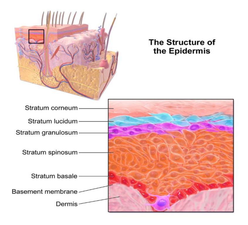
Figure 5.9 Structure of the Epidermis.
Epidermal Layers: the layers or strata progression from the superficial to the deep layers.
5.19 Characteristics of this Layer:
- Mostly keratinocytes
- reproductive actively mitotic division
- produce keratin with cell progression upward keratinization.
5.19.1 Strata of the Epidermis:
- superficial stratum stratum corneum
- intermediate stratum
- stratum lucidum absent in “thin” skin.
- stratum granulosum
- stratum spinosum
- bottom stratum stratum basale (germinitivum)
5.20 Skin Pigmentation
Melanocytes. Melanocytes are located in the stratum basale as can be noted in Figures 5.10 & 5.11.
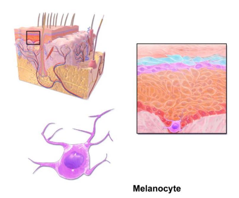
Figure 5.10 Epidermis with Melanocyte.
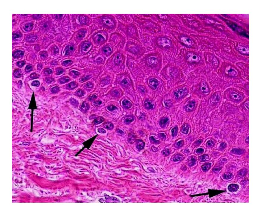
Figure 5.11 Examples of Melanocytes in the Basal Layer of the Epidermis.
5.21 Dermal and Subcutaneous Layers
5.22 Layers of the Dermis
- Papillary layer
- Reticular layer
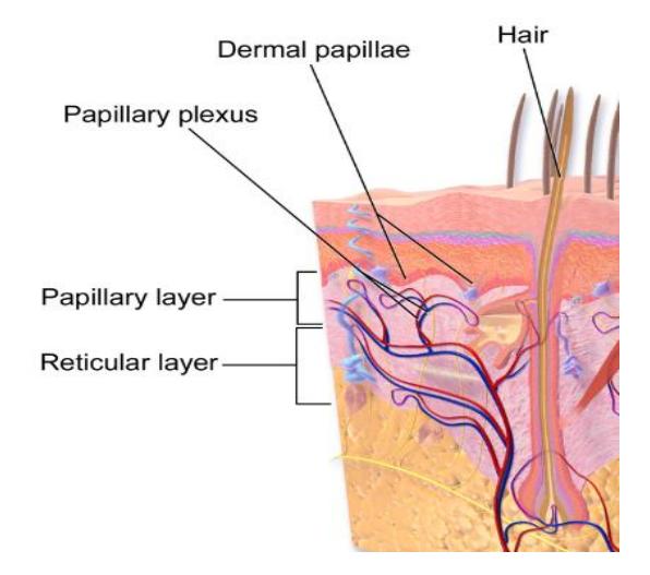
Figure 5.12 Layers of the dermis.
5.23 Hypodermis (Subcutaneous)
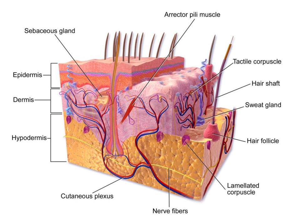
Figure 5.13 Anatomy of the Skin.
5.24 Dermal and Hypodermal Structures
Using a model or diagram of the integumentary system, identify and describe the function of the following integumentary structures:
- Dermal papillae
- Hair shaft
- Hair root
- Hair follicle
- Pore
- Arrector pili muscle
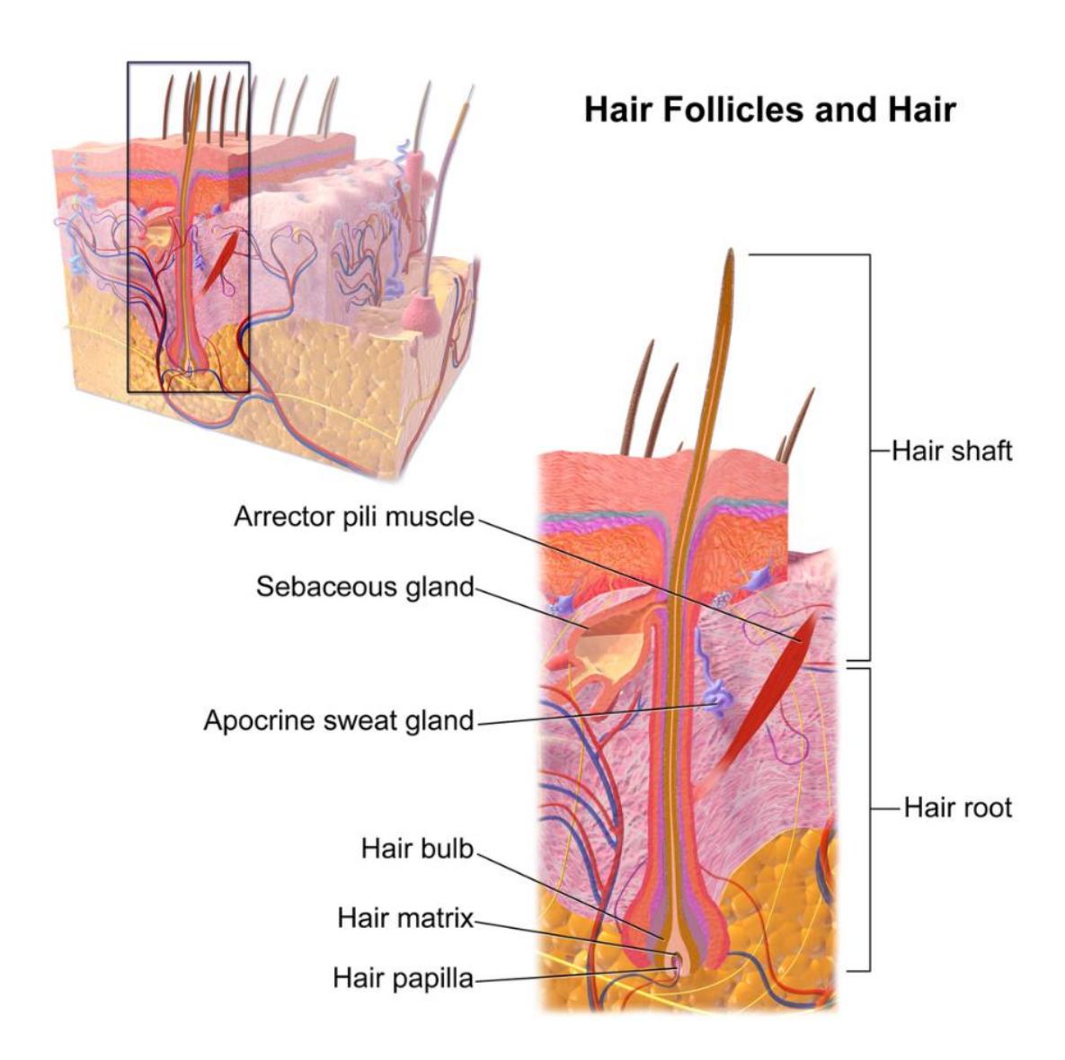
Figure 5.14.
Figure 5.14 Hair Follicle.
5.25 Post Lab Activities
5.26 Test your recall:
5.27 Post Lab Activity 5.1: Answer the concept questions
- Name the layers of the dermis.
Papillary layer; Reticular layer
- List three major types of glands associated with the integumentary system and describe their function.
Sebaceous glands – secrete sebum to lubricate skin Merocrine (eccrine) glands – regulate temperature via sweat Apocrine glands – produce thicker sweat in specific regions
- What are the three protective functions of the integumentary system?
Protection from pathogens Prevention of water loss Protection from UV radiation
5.28 Post Lab Activity 5.2
5.29 Matching
5.30 Table 5.4 Matching: Write the Letter of the Definition in the Column to the Left of the Term
| # | Terms | Definitions |
|---|---|---|
| 1 | Pore (e) | a. The part of a hair projecting beyond the surface of the skin |
| 2 | Arrector pili muscle (h) | b. An onion-shaped structure that is responsible for sensitivity to vibration and pressure |
| 3 | Meissner’s corpuscle (j)’s corpuscle | c. Any of the small sacculated glands lodged in the substance of the derma, usually opening into the hair follicles, and secreting an oily material |
| 4 | Pacinian corpuscle (g) corpuscle | d. An organ found in mammalian skin which regulates hair growth |
| 5 | Keratinocyte (k) | e. A small opening or empty space. |
| 6 | Apocrine sweat gland (i) | f. The enlarged basal part of a hair within the skin |
| 7 | Hair follicle (d) | g. A type of nerve ending in the skin that is responsible for sensitivity to pressure |
| 8 | Dermal papillae (m) | h. The small smooth muscles associated with the base of each hair that contract when the body surface is chilled and erect the hairs |
| 9 | Merocrine sweat gland (l) | i. Glands found in places like the armpits, scrotum, and anus. They tend to open into hair follicles instead of hairless areas of skin. |
| 10 | Hair root (f) | j. An unspecialized, afferent nerve fiber ending of a sensory neuron |
| 11 | Sebaceous gland (c) | k. An epidermal cell that produces keratin. |
| 12 | Free nerve ending (j) | l. True sweat glands in the sense of helping to regulate body temperature |
| 13 | Hair shaft (a) | m. Any vascular protuberance of the skin’s dermal layer extending into the epidermal layer, often containing tactile corpuscles |
Match the following terms with the correct parts in figure 5.15.
Table 5.5 Identification of the Dermal Structures
| Dermal Structure | Number on Figure 5.15 |
|---|---|
| Pore | |
| Arrector pili muscle | |
| Meissner’s corpuscle | |
| Pacinian corpuscle | |
| Keratinocyte | |
| Apocrine sweat gland | |
| Hair follicle | |
| Dermal papillae | |
| Merocrine sweat gland | |
| Hair root | |
| Sebaceous gland | |
| Free nerve ending | |
| Hair shaft |
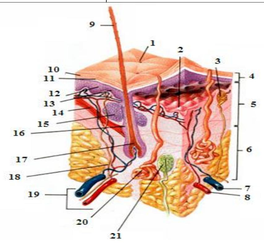
Figure 5.15 Anatomy of Human Skin Sans Labels.
Match the statement blank with the correct answer.
Table 5.6 Match the statement with the best answer.
| Answer | Statement | Possible Answer |
|---|---|---|
| a. | The epidermis is composed of __________. | Hair bulb |
| b. | The deepest layer of the epidermis. | Sudoriferous |
| c. | The majority of the cells of the epidermis are __________ , so-called because they make __________. | stratum basale |
| d. | Melanocytes are located in the __________ (which layer?). | stratum lucidum |
| e. | Immune cells in the stratum spinosum which protect against invaders are the __________. | root |
| f. | A very thin layer called the __________ is in the epidermis but is visible only in thick skin. | keratin |
| g. | The first level of protection against abrasion and toxic chemicals at the body’s surface is provided by the __________. | Hair shaft |
| h. | The dermis consists of two layers: the __________, which is characterized by a dimpled interface with the epidermis, and the __________, which accounts for 80% of the skin’s thickness | Pacinian corpuscles; |
| i. | The reticular layer of the dermis contains __________ to sense firm pressure. | papillary layer; reticular layer |
| j. | Apocrine and eccrine glands are both __________ (sweat) glands. | keratinocytes; keratin |
| k. | __________ glands produce a waxy substance to protect the ear canal from dust, small insects, etc. | squamous epithelial tissue |
| l. | The portion of the hair that is above the surface of the skin is called the __________. | Stratum corneum |
| m. | The sub-surface portion of the hair is called the __________. | Sudoriferous |
| n. | The origin of the hair shaft, deep within the dermis, is called the __________. | Ceruminous |
| o. | The sheath in which a hair is held is called __________. | Langerhans’ cells |
| follicle |
5.31 Post Lab Activity 5.3
Crossword Puzzle: Complete the following crossword puzzle.
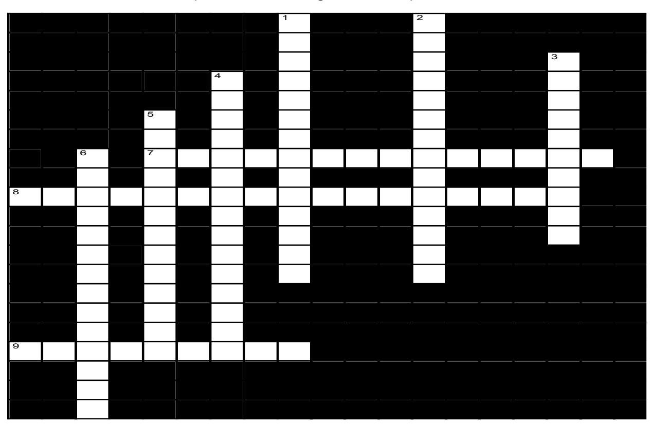
Figure 5.16 The Integumentary System
5.31.1 Across
- 7 the layer which serves to strengthen the skin and provides our skin with elasticity. It contains hair follicles, sweat glands, and sebaceous glands. (9,5)
- 8 a thin layer of cells in the epidermis lying above the stratum spinosum and below the stratum corneum. (7,10)
- 9 the outermost of the three layers that comprise the skin (9)
5.31.2 Down
1 the outermost layer of the epidermis comprised of several levels of flattened corneocytes (7,7)
2 a more superficial and significantly thinner layer of the dermis, mainly composed of areolar connective tissue. (9,5)
3 the lowermost layer of the integumentary system in vertebrates. (10)
4 a layer of the epidermis composed of polyhedral keratinocytes. (7,8)
5 the deepest layer of the five layers of the epidermis, the external covering of skin in mammals. The stratum. It is a single layer of columnar or cuboidal basal cells (7,6)
6 a thin, clear layer of dead skin cells in the epidermis named for its translucent appearance only in areas of thick skin (7,7)
5.32 Post Lab Activity 5.4
5.33 Check Your Understanding
- You are a medical examiner (ME) who is performing an autopsy on a shooting victim. The bullet did not pass through the body and was lodged in the abdomen near the spine. The ME must find and retrieve the bullet to determine its caliber, so she needs to cut through the integument. List the layers that you would cut through from the superficial to the deepest layer.
- It is late in the summer and a young couple that had planted a garden began to freeze and can produce their products for use later in the year. Their garden was very productive, so they were able to store carrots, corn, sweet potatoes, spinach, butternut squash, broccoli, and cantaloupe for use in the winter. They have a four-month-old daughter, and they noticed that after they began feeding her some of the canned food her skin developed a yellowish orange tint. They immediately took her to the general practitioner (GP), and he checked out the child. Following the examination, he returned the child to the parents and with a smirk and gleam in his eyes he indicated that the child was fine but cautioned them that they should vary her diet. Explain why the GP is not concerned about the child’s skin condition?
- Discuss how the Integumentary system’s skin helps to regulate your body temperature, when your body temperature rises above normal.
- How would you explain why a light skinned individual would be more susceptible to malignant melanoma?
5.34 Post Lab Activity 5.5
5.35 Test Your Recall
Label the layers and structures of the epidermis.
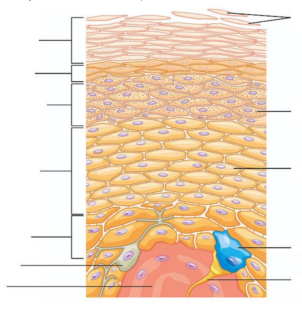
Figure 5.17 Layers of the Epidermis.
Using the picture below, identify the substructures within the following accessory structures.
- Hair shaft
- Hair root
- Hair bulb
- Hair follicle
- Arrector pili muscle
- Sebaceous gland
- Apocrine sweat gland
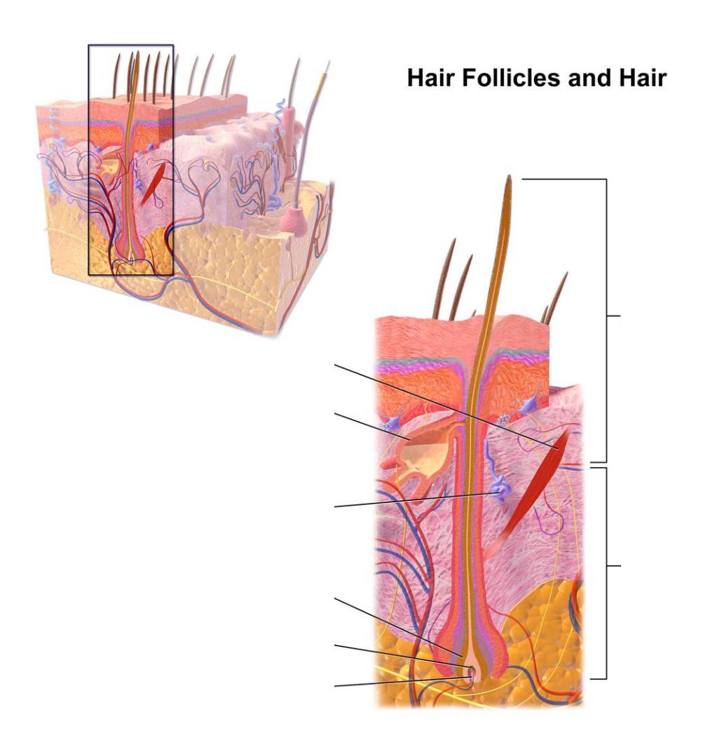
Figure 5.18 Illustration from Anatomy & Physiology of the Epidermis.
5.36 Post Lab Activity 5.6
5.37 Fill in the blanks
- Name the layers of the dermis.
Papillary layer; Reticular layer
Sebaceous glands – secrete sebum to lubricate skin Merocrine (eccrine) glands – regulate temperature via sweat Apocrine glands – produce thicker sweat in specific regions | |——|——————————————————–| | a) | | | b) | | | c) | |
Protection from pathogens Prevention of water loss Protection from UV radiation | |——|——————————————————–| | a) | | | b) | | | c) | |
| List four functions of the integumentary system | |
|---|---|
| a) | |
| b) | |
| c) | |
| d) |
- The skin is composed of what type of epithelial tissue?
| 1) | List the five layers of the epidermis found in thick skin. Indicate which layer is not present in thin skin. |
|---|---|
| a) | |
| b) | |
| c) | |
| d) | |
| e) |
| 2) | Identify four accessory structures found in the dermis, describe their function. |
|---|---|
| a) | |
| b) | |
| c) | |
| d) |
5.38 Answer keys
5.38.0.1 Crossword Puzzle
Across: 7 Reticular layer, 8 Stratum granulosum, 9 Epidermis.
Down: 1 Stratum corneum, 2 Papillary layer, 3 Hypodermis, 4 Stratum spinosum, 5 Stratum basale, 6 Stratum lucidum.
Chapter 5: Integumentary System
| Key Terms | Definitions |
|---|---|
| acne | skin condition due to infected sebaceous glands |
| albinism | genetic disorder that affects the skin, in which there is no melanin production |
| apocrine sweat gland | sweat gland that is armpits and genital regions |
| arrector pili | smooth muscle that is activated in response to external stimuli that pull on hair follicles and make the hair “stand-up” |
| basal cell | type of stem cell found in the stratum basale and in the hair matrix that continually undergoes cell division, producing the keratinocytes of the epidermis |
| basal cell carcinoma | cancer that originates from basal cells in the epidermis of the skin |
| bedsore | sore on the skin that develops when regions of the body start necrotizing due to constant pressure and lack of blood supply; also called decubitus ulcers |
| callus | thickened area of skin that arises due to constant abrasion |
| corn | type of callus that is named for its shape and the elliptical motion of the abrasive force |
| cortex | in hair, the second or middle layer of keratinocytes originating from the hair matrix, as seen in a cross-section of the hair bulb |
| cuticle | in hair, the outermost layer of keratinocytes originating from the hair matrix, as seen in a cross section of the hair bulb |
| dermal papilla (plural = dermal papillae) | extension of the papillary layer of the dermis that increases surface contact between the epidermis and dermis |
| dermis | layer of skin between the epidermis and hypodermis, composed mainly of connective tissue and containing blood vessels, hair follicles, sweat glands, and other structures |
| desmosome | structure that forms an impermeable junction between cells |
| eccrine sweat gland | sweat gland that is common throughout the skin surface; it produces a hypotonic sweat for thermoregulation |
| eczema | skin condition due to an allergic reaction, which resembles a rash |
| elastin fibers | fibers made of the protein elastin that increase the elasticity of the dermis |
| epidermis | outermost tissue layer of the skin |
| external root | outer layer of the hair follicle that is an extension of the epidermis, which |
| sheath | encloses the hair root |
| first-degree burn | superficial burn that injures only the epidermis |
| fourth-degree burn | burn in which full thickness of the skin and underlying muscle and bone is damaged |
| free nerve ending | a.k.a. bare nerve ending, is an unspecialized, afferent nerve fiber sending its signal to a sensory neuron. |
| hair | keratinous filament growing out of the epidermis |
| hair bulb | structure at the base of the hair root that surrounds the dermal papilla |
| hair follicle | cavity or sac from which hair originates |
| hair matrix | layer of basal cells from which a strand of hair grows |
| hair papilla | mass of connective tissue, blood capillaries, and nerve endings at the base of the hair follicle |
| hair root | part of hair that is below the epidermis anchored to the follicle |
| hair shaft | part of hair that is above the epidermis but is not anchored to the follicle |
| hypodermis | connective tissue connecting the integument to the underlying bone and muscle |
| hyponychium | thickened layer of stratum corneum that lies below the free edge of the nail |
| integumentary system | skin and its accessory structures |
| internal root sheath | innermost layer of keratinocytes in the hair follicle that surround the hair root up to the hair shaft |
| keloid scar | scar that has layers raised above the skin surface |
| keratin | structural protein that gives skin, hair, and nails its hard, water-resistant properties |
| keratinocyte | cell that produces keratin and is the most predominant type of cell found in the epidermis |
| Langerhans cell | specialized dendritic cell found in the stratum spinosum that functions as a macrophage |
| lunula layer of thick epithelium | basal part of the nail body that consists of a crescent-shaped layer of thick epithelium |
| medulla | in hair, the innermost layer of keratinocytes originating from the hair matrix |
| Meissner corpuscle (also, tactile corpuscle) | in the skin that responds to light touch |
| melanin | pigment that determines the color of hair and skin |
| melanocyte | cell found in the stratum basale of the epidermis that produces the pigment melanin |
| melanoma | type of skin cancer that originates from the melanocytes of the skin |
| melanosome | intercellular vesicle that transfers melanin from melanocytes into keratinocytes of the epidermis |
| merocrine gland | See eccrine gland |
| Merkel cell | receptor cell in the stratum basale of the epidermis that responds to the sense of touch |
| metastasis | spread of cancer cells from a source to other parts of the body |
| nail bed | layer of epidermis upon which the nail body forms |
| nail body | main keratinous plate that forms the nail |
| nail cuticle | fold of epithelium that extends over the nail bed, also called the eponychium |
| nail fold | fold of epithelium at that extend over the sides of the nail body, holding it in place nail root part of the nail that is lodged deep in the epidermis from which the nail grows |
| Pacinian corpuscle (also, lamellated corpuscle) | receptor in the skin that responds to vibration |
| papillary layer | superficial layer of the dermis, made of loose, areolar connective tissue |
| reticular layer | deeper layer of the dermis; it has a reticulated appearance due to the presence of abundant collagen and elastin fibers |
| rickets | disease in children caused by vitamin D deficiency, which leads to the weakening of bones |
| scar | collagen-rich skin formed after the process of wound healing that is different from normal skin |
| sebaceous gland | type of oil gland found in the dermis all over the body and helps to lubricate and waterproof the skin and hair by secreting sebum |
| sebum | oily substance that is composed of a mixture of lipids that lubricates the skin and hair |
| second-degree burn | partial thickness burn that injures the epidermis and a portion of the dermis |
| squamous cell | type of skin cancer that originates from the stratum spinosum of the |
| carcinoma | epidermis |
| stratum basale | deepest layer of the epidermis, made of epidermal stem cells |
| stratum corneum | most superficial layer of the epidermis |
| stratum granulosum | layer of the epidermis superficial to the stratum spinosum |
| stratum lucidum | layer of the epidermis between the stratum granulosum and stratum corneum, found only in thick skin covering the palms, soles of the feet, and digits |
| stratum spinosum | layer of the epidermis superficial to the stratum basale, characterized by the presence of desmosomes |
| sudoriferous gland | sweat gland |
| third-degree burn | burn that penetrates and destroys the full thickness of the skin (epidermis and dermis) |
| vitamin D | compound that aids absorption of calcium and phosphates in the intestine to improve bone health |
| vitiligo | skin condition in which melanocytes in certain areas lose the ability to produce melanin, possibly due an autoimmune reaction that leads to loss of color in patches |